What is a dental film?
A targeted small X-ray, now often digital.

A periapical dental X-ray targets a small area. It can show one or more teeth with their roots and surrounding bone. The term “dental film” is still used even for digital images. Whether an image is needed depends on examination findings and the specific diagnostic question.
For dental trainees & teams
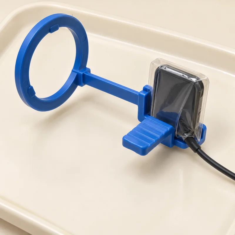
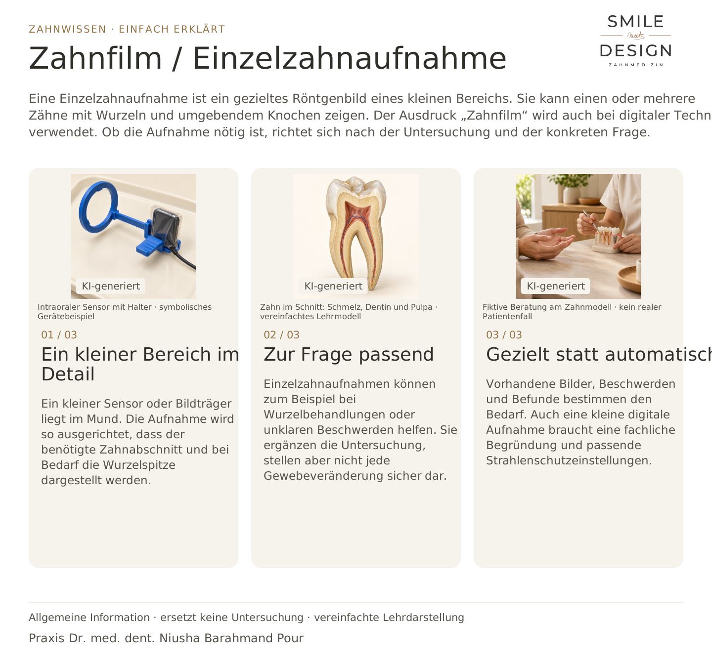
A small sensor or receptor is placed inside the mouth. Positioning aims to show the required tooth area and, when needed, its root tip.
Periapical images can help with root canal treatment or unexplained symptoms. They complement examination but do not reveal every tissue change reliably.
Existing images, symptoms and findings determine the need. Even a small digital X-ray requires justification and appropriate radiation-protection settings.
A targeted small X-ray, now often digital.
Selected questions about teeth, roots and surrounding bone.
Examination, symptoms, existing images and expected diagnostic benefit.
Three steps. Straight to the point.
Explanations are also shown on screen.

Silent film in German · English explanations in the three views above
A photograph shows visible surfaces without X-ray radiation. A dental X-ray uses ionising radiation to depict internal hard structures. They answer different questions.
A panoramic image provides an overview but has overlapping structures and limits to detail. A targeted image may be better for particular root or bone questions.
Bitewings mainly show the crowns of neighbouring upper and lower teeth. A periapical image aims to show a tooth down to its root tip and surrounding tissues.
No. X-rays mainly show changes in hard structures. Early inflammation may have no clear radiographic sign; symptoms and other tests remain important.
A question to discuss with Dr. med. dent. Niusha Barahmand Pour:
What additional information would a periapical image provide about this tooth?
Symptoms, examination findings and personal wishes form the basis of individual treatment planning.
An image helps when it answers a specific question about the tooth.
Discuss an unclear dental findingChoose an appointment on DoctolibBuild on the simple explanation with clinical terms, key distinctions and a knowledge question.
Even a detailed two-dimensional image can contain overlapping structures. Absence of radiographic signs does not always exclude early disease.
Editorial research: 23 October 2026. This explanation covers general principles and does not replace a personal examination. Suitable treatment is discussed following individual assessment.