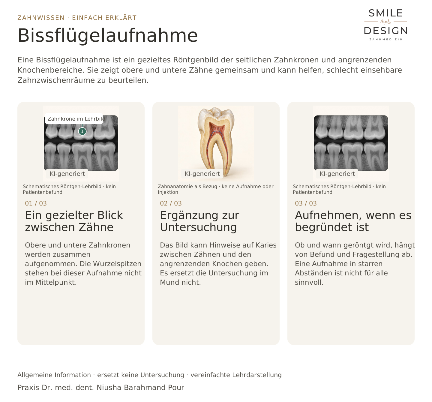
What does the image show?
Upper and lower back tooth crowns, the spaces between them and nearby bone.

A bitewing is a targeted X-ray of the crowns of back teeth and nearby bone. It shows upper and lower teeth together and can help assess areas between teeth that are difficult to see.
For dental trainees & teams
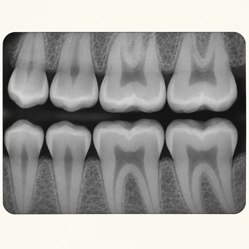
Upper and lower tooth crowns appear together. The root tips are not the main focus of this image.
The image can provide information about decay between teeth and nearby bone. It does not replace examination of the mouth.
Whether and when an X-ray is needed depends on the findings and clinical question. Fixed intervals do not suit everyone.
Upper and lower back tooth crowns, the spaces between them and nearby bone.
When a specific question requires information beyond the clinical examination.
No. The need for imaging and repeat images is justified individually.
Three steps. Straight to the point.
Explanations are also shown on screen.

Silent film in German · English explanations in the three views above
A bitewing shows a targeted area. A panoramic image offers a wider overview, but not the same detail.
No. It is limited to selected tooth crowns, spaces between them and nearby bone.
No. X-rays need professional interpretation alongside the clinical findings.
Radiation is used when the diagnostic benefit is justified. Existing images and individual risks also matter.
A question to discuss with Dr. med. dent. Niusha Barahmand Pour:
What specific question should this X-ray answer, and could existing images help?
The prerequisites are assessed before selecting a treatment.
Questions about necessary examinations belong in individual planning.
Discuss the examinationChoose an appointment on DoctolibBuild on the simple explanation with clinical terms, key distinctions and a knowledge question.
A bitewing shows a targeted area, not a complete view of every root tip.
Editorial research: 23 October 2026. This explanation covers general principles and does not replace a personal examination. Suitable treatment is discussed following individual assessment.