
Dental insights · New developments
Digital impressions: What an intraoral scan can — and cannot — do.
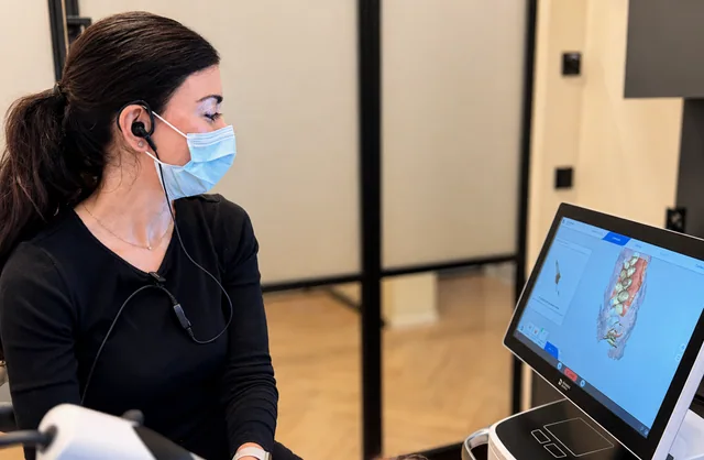
Instead of a filled impression tray, a small handheld device is moved along the teeth. A three-dimensional model of the mouth appears on screen. This is called an intraoral scan or digital impression. The technology can support treatment steps and make explanations clearer. However, a digital model does not mean that every examination can now be carried out on a computer.
Editorial review date:
What is recorded
An intraoral scanner records the visible surfaces of teeth and gums and combines the images into a digital model. University College London Hospital describes scanning as an alternative to conventional impressions made with impression material. The model can be viewed immediately on screen and used for further treatment. UCLH: Digital impressions in dentistry
For patients, a particularly helpful feature is often being able to discuss their own situation together on screen. They no longer have to rely solely on imagining which tooth surface or gap is being discussed. However, the model remains a record: examination, findings and consultation establish which treatment is appropriate.
What may be different at the appointment
Scanning avoids having impression material in the mouth for this recording. An NHS hospital report from Somerset also describes how scanning can be interrupted for a break. Whether this feels more comfortable also depends on the individual situation. If mouth opening or gagging concerns you, mention it before the scan. Somerset NHS: Experiences with digital impressions
This does not promise that every appointment will be shorter or that every conventional impression can be avoided. A more useful specific question is: “What is this scan needed for in my case, and what further steps follow?” This helps put its benefit into context better than the word “digital” alone.
What the new guideline states
The first S2k guideline on intraoral scanning in dentistry was published in March 2026. It brings together professional recommendations on requirements and uses. Its evidence review dates back to 2025; the publication year is not the date when every individual recommendation originated. AWMF: Guideline publication
The guideline lists digital models for planning and monitoring progress as possible applications. For a useful result, the relevant surfaces must be clearly visible and accessible; moisture and other recording conditions also play a role. Digital working records are subject to the same quality requirements as conventional impressions. AWMF/DGZMK/DGCZ: S2k guideline on intraoral scanning
Where the limitations lie
According to the guideline, an intraoral scan does not replace conventional caries diagnosis using clinical examination and, where appropriate, X-rays. Nor does a tooth colour displayed on screen currently replace conventional shade selection in general. The entire processing chain also matters for the eventual treatment result, not just the scan itself. S2k guideline: Diagnosis, shade selection and further processing
Three questions to help put it into context
Ask what the scan is intended to answer for your specific treatment, which additional information is needed and whether you can look at the model together. Also ask whether a screen image shows the current findings or an initial treatment proposal. These communicate different things. Digital technology is particularly helpful when it improves your understanding of the decision — not simply because a device looks new.
Technology should be understandable.
It should be clear what information an examination or scan provides. Read more about our approach to personal conversation in our philosophy.
Get in touchProfessional sources
Research and editorial review: 23 September 2026. This information provides general guidance. An individual examination and consultation establish which treatment suits your situation.