
Dental insights · Did you know?
White spots: Is every white mark tooth decay?
A pale spot on an incisor often first catches your eye in a photograph. Sometimes it remains after fixed braces; sometimes it has been present since the tooth erupted. “White spot” initially describes its appearance — not its cause. This distinction determines whether prevention, targeted treatment or primarily an aesthetic consultation is appropriate.
Editorial review date:
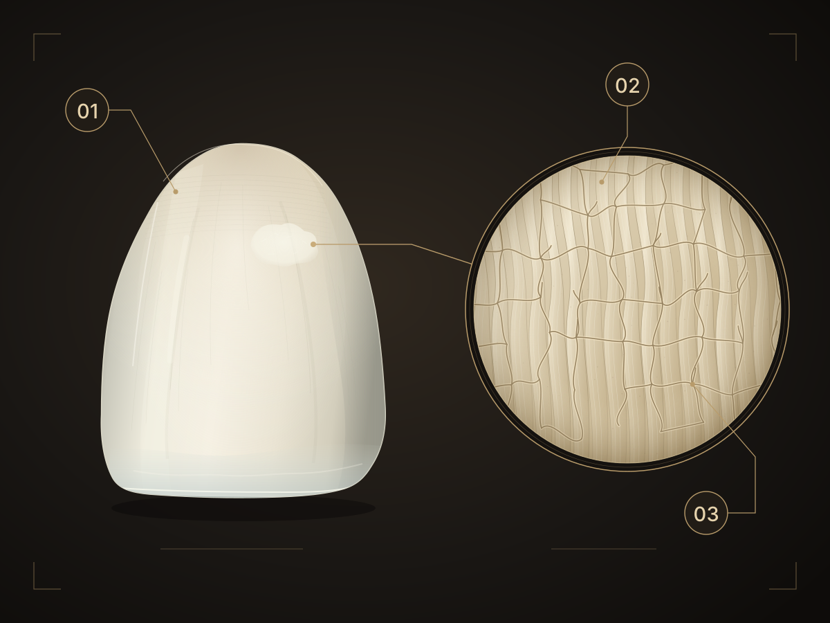
White does not automatically mean decay.
White changes may result from mineral loss in early decay. There are also enamel opacities: areas that formed differently during tooth development. Examples include fluorosis and certain mineralisation disorders. Colour or shape alone cannot reliably identify the cause. NIDCR: The development of decay and fluorosis
Molar-incisor hypomineralisation, or MIH, can affect the incisors as well as the first permanent molars. A single pale spot on a front tooth is not enough for this diagnosis. The full dentition and any symptoms are assessed. EAPD: Criteria for MIH
Early decay: the process before a cavity.
In tooth decay, bacteria in dental plaque produce acids from sugar. If more minerals are lost over time than are replaced, the enamel changes. An early lesion may look whitish even though the surface has not yet broken down. At this stage, the process can be arrested under suitable conditions; fluoride and minerals from saliva support remineralisation. However, an existing cavity does not grow back in this way. NIDCR: Early decay and cavities
Disease activity and visibility are not the same: even an arrested lesion may remain visible as a spot. Before making a cosmetic change, we therefore clarify whether active decay needs treatment. Clinical study of inactive white spots
What does infiltration do?
During resin infiltration, the surface is prepared and a low-viscosity material is introduced into accessible porous areas of the enamel and light-cured. This may reduce the visual contrast of a white area. The method does not build new enamel and is not whitening.
In a clinical study of decay-related white spots during treatment with fixed braces, the process included cleaning, gum protection, isolation, etching, infiltration and polishing. Etching makes the surface accessible to the material; “microinvasive” therefore does not mean “without any removal of tooth structure”. The precise application depends on the findings and the intended material. Kashash et al.: Treatment process and optical principle
Why is not every spot suitable?
Key factors include the cause, the depth of the change and the condition of the surface. Results for decay-related white spots after braces do not establish general suitability for developmental opacities. Evidence for aesthetic infiltration in MIH is limited. Surfaces that have already broken down need a different treatment plan. EAPD: Options and limitations in MIH
Known material allergies and the ability to keep the tooth sufficiently dry and protect the gums also inform individual treatment planning. The intended material is checked accordingly. Complete disappearance of a spot cannot be promised; nor can the duration of the aesthetic result be predicted for every tooth.
What do studies support?
A randomised study of white spots after fixed braces found greater aesthetic improvement with infiltration than in the untreated comparison group. Follow-up lasted eight weeks. This supports a possible masking effect in the situation studied, but not a guaranteed lifelong result for every white spot. Senestraro and colleagues: Clinical study
This is distinct from controlling decay between teeth: in another randomised study, selected early, non-cavitated lesions progressed less often after infiltration over an average of seven years. All participants also received advice on diet, interdental cleaning and fluoride. These findings do not address colour matching in front teeth. Paris and colleagues: Seven-year data
Three questions for your appointment.
- What is the cause? Early decay and a developmental opacity do not require the same treatment plan.
- What is the aim? Controlling decay, changing appearance — or both?
- What remains important afterwards? Ask for an explanation of care, fluoride use and review intervals suited to your situation.
Infiltration does not replace prevention. Equally, a visible spot does not automatically mean immediate treatment is needed. An examination helps distinguish necessary care from a personal aesthetic choice.
Understand the cause first.
Our page on white spot treatment explains the options, process and limitations of resin infiltration.
Get in touchProfessional sources
Research and editorial review: 24 September 2026. General guidance; diagnosis and individual treatment planning require a dental examination. Clinical studies relate to the specific patient groups, materials and treatment protocols studied. Their findings cannot be applied universally to every white spot.
- NIDCR: The decay process, early lesions and fluorosis – public patient information, updated January 2025.
- EAPD: European guidance on MIH – updated policy document, 2022.
- Kashash et al.: Infiltration of decay-related white spots during fixed appliance treatment – randomised study, Clinical Oral Investigations 2024, six-month follow-up.
- Wang et al.: Comparison of four treatments for white spots after orthodontics – randomised study, Clinical Oral Investigations 2023, twelve-month follow-up.
- ADA: Guideline on nonrestorative treatments for carious lesions – 2018; treatment options depend on factors including the tooth surface and findings.
- Senestraro et al.: Infiltration of inactive white spots after orthodontics – randomised study, JADA 2013.
- Paris et al.: Seven-year observation of infiltrated proximal caries – randomised study, Journal of Dentistry 2020.