What is CBCT?
An X-ray examination used to calculate a three-dimensional dataset.

Cone beam CT, known as DVT in German, produces three-dimensional X-ray images of teeth and bony structures. It may be useful for selected questions when additional information beyond conventional images is needed.
For dental trainees & teams
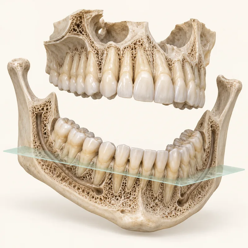
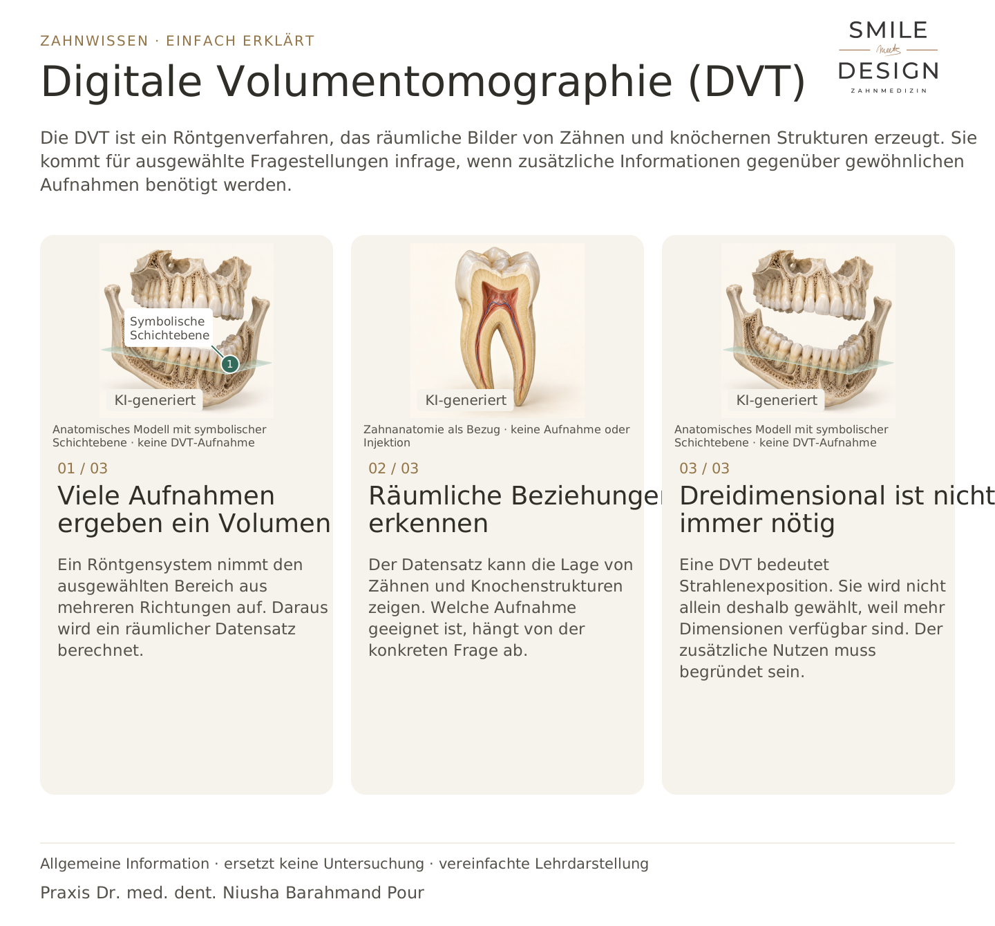
An X-ray system records a selected area from different directions. These images are used to calculate a three-dimensional dataset.
The dataset can show the position of teeth and bony structures. The appropriate type of image depends on the clinical question.
CBCT involves radiation exposure. It is not chosen simply because more dimensions are available. Its additional benefit needs justification.
An X-ray examination used to calculate a three-dimensional dataset.
For selected questions that require additional spatial information.
The potential diagnostic benefit is weighed against radiation exposure.
Three steps. Straight to the point.
Explanations are also shown on screen.

Silent film in German · English explanations in the three views above
CBCT produces X-ray images of a volume. An intraoral scan optically records accessible surfaces.
No. It uses X-rays, unlike an optical intraoral scan.
Not automatically. Imaging is chosen according to the anatomy and the information needed for planning.
No. It is mainly suited to hard structures and replaces neither clinical examination nor every other imaging method.
A question to discuss with Dr. med. dent. Niusha Barahmand Pour:
What additional information would we need, and which examination is suitable?
The profile describes digital planning as a support for personal consultation.
The clinical question helps determine which imaging may be useful.
Discuss imagingChoose an appointment on DoctolibBuild on the simple explanation with clinical terms, key distinctions and a knowledge question.
A three-dimensional image is not automatically diagnostically better; it should add useful information for the clinical question.
Editorial research: 23 October 2026. This explanation covers general principles and does not replace a personal examination. Suitable treatment is discussed following individual assessment.