No X-rays?
Correct.

MRI can show soft tissues of the temporomandibular joint, particularly the articular disc and joint fluid. It uses magnetic fields rather than X-rays. Whether it can answer a useful question is decided after history and examination; not every click requires MRI.
For training & practice ↓
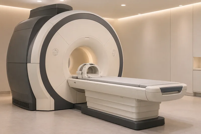
The position or shape of the disc may be relevant. Imaging is selected when it can contribute to assessment.
Depending on the question, images may be obtained in different mouth positions to compare joint relationships.
An unusual image does not automatically explain every symptom. Imaging and clinical findings are considered together.
The position or shape of the disc may be relevant. Imaging is selected when it can contribute to assessment.
Depending on the question, images may be obtained in different mouth positions to compare joint relationships.
An unusual image does not automatically explain every symptom. Imaging and clinical findings are considered together.
Correct.
No.
Treating professionals and radiology.
Three steps. No audio.
Each scene can also be selected manually.

Silent educational film with German on-screen text and simplified teaching images. Not a patient case or a promised treatment outcome. The full English explanation is available as text.
Soft-tissue and bone questions do not always require the same imaging method. More imaging does not automatically mean more benefit.
No. MRI is useful for particular soft tissues. CBCT uses X-rays and principally depicts bony structures.
No. Symptoms, function, examination and the specific question guide imaging. A sound alone does not determine the investigation.
Implants, electronic devices in the body, possible metal fragments and claustrophobia should be discussed during radiology safety screening. Do not assess MRI compatibility yourself.
Editorially suggested question to discuss with Dr Niusha Barahmand Pour:
What specific question would additional imaging answer in my case?
Jaw discomfort or limited movement? First establish which examination would be useful.
Discuss jaw symptomsAppointment selection at DoctolibSoft-tissue and bone questions do not always require the same imaging method. More imaging does not automatically mean more benefit.
Editorial research: . General information, not an individual diagnosis or treatment recommendation. Teaching illustrations are simplified and do not depict a patient case.