What is it?
An optical tool.

A dental operating microscope magnifies the treatment field and supports directed illumination. It can help reveal fine structures such as root canal entrances. It is a tool for the clinician, not a separate treatment.
For training & practice ↓
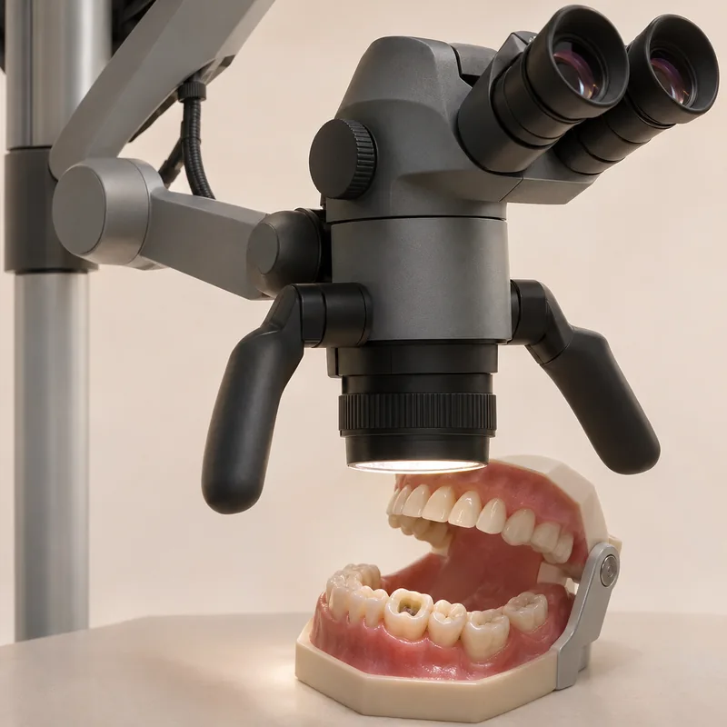
Fine structures appear larger. Usefulness also depends on access, line of sight and operator experience.
In endodontics it may help assess canal entrances or some obstructions. It cannot see around every curve in a canal.
Diagnosis, cleaning, sealing and follow-up remain important. A microscope does not promise painless care or a particular outcome.
An optical tool.
For selected diagnostic and treatment steps involving fine structures.
What benefit is expected for this tooth and what limits remain.
Three steps. No audio.
Each scene can also be selected manually.

Silent educational film with German on-screen text and simplified teaching images. Not a patient case or a promised treatment outcome. The full English explanation is available below.
Fine structures appear larger. Usefulness also depends on access, line of sight and operator experience.
In endodontics it may help assess canal entrances or some obstructions. It cannot see around every curve in a canal.
Diagnosis, cleaning, sealing and follow-up remain important. A microscope does not promise painless care or a particular outcome.
Both provide optical magnification. Construction, magnification options and lighting differ; neither replaces clinical judgement.
No. Optical inspection and imaging answer different questions.
No. A scan records surfaces digitally; a microscope magnifies the visible working field.
Appropriate magnification and other aids depend on the findings and complexity of the procedure. The treatment discussion can clarify which aids are planned and what they are intended to contribute for this tooth.
An editorially suggested question for a discussion with Dr med dent Niusha Barahmand Pour:
Which tools are appropriate for this tooth, and why?
Before root canal treatment, a clear plan helps: ask about the purpose and limitations of proposed tools.
Discuss the treatment planAppointment selection at DoctolibBoth provide optical magnification. Construction, magnification options and lighting differ; neither replaces clinical judgement.
Editorial research: . General information, not an individual diagnosis or treatment recommendation. Teaching illustrations are simplified and do not depict a patient case.