Where is it?
At the roof of the mouth.

The palate forms the roof of the mouth. Its front section is the bone-supported hard palate; its back section is the mobile soft palate, also called the velum. The hard palate separates the oral and nasal spaces. The soft palate contributes to swallowing and speech. These parts belong together but have different structures and roles.
For training & practice ↓
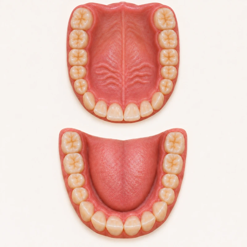
Mucosa covers the bone-supported hard palate. It forms the front of the mouth roof; the visible ridges are surface landmarks.
The soft palate contains muscle and changes position during swallowing and speech. The uvula hangs from its free back edge.
Hard or soft, right or left: location helps the examination. A new swelling or persistent change in the lining needs separate assessment.
Mucosa covers the bone-supported hard palate. It forms the front of the mouth roof; the visible ridges are surface landmarks.
The soft palate contains muscle and changes position during swallowing and speech. The uvula hangs from its free back edge.
Hard or soft, right or left: location helps the examination. A new swelling or persistent change in the lining needs separate assessment.
At the roof of the mouth.
No; its back section is soft.
No.
Three steps. No audio.
Each scene can also be selected manually.

Silent educational film with German on-screen text and simplified teaching images. Not a patient case or a promised treatment outcome. The full English explanation is available as text.
Palate names the structure. Palatal describes a position or direction towards it, such as the inner surface of an upper tooth.
No. The maxilla is a bone. The palate includes a front section supported by bone and a soft back section, both covered by mucosa.
Yes. It hangs from the back edge of the soft palate but is not the entire soft palate.
Palatal rugae are normal surface features of the anterior palatal lining. A new painful change should be assessed separately.
A photograph can show location. It cannot reliably assess movement or coordination during swallowing and speech.
New lumps, persistent red or white patches, and an ulcer lasting more than three weeks should be examined. There are many possible causes; a photograph cannot establish the diagnosis.
You can bring this question to your appointment:
Does my finding involve the hard or soft palate, and what assessment is appropriate?
A dental examination can clarify how this relates to your findings and treatment planning.
Discuss your dental findingsAppointment selection at DoctolibPalate names the structure. Palatal describes a position or direction towards it, such as the inner surface of an upper tooth.
Editorial research: . General information, not an individual diagnosis or treatment recommendation. Teaching illustrations are simplified and do not depict a patient case.
If you turn this on, this website stores which guides you open, separately from advertising preferences. The markers expire after a fixed 30-day period from activation. The list is not sent to us by this feature. You can turn it off here and delete the markers.
Other people using the same browser profile can also see the markers. German and English versions share one marker. Expired entries are removed the next time you open a page with this feature; deleting browser data may remove them earlier. “Viewed” means opened, not read or completed.