What identifies the cyst?
Assessment of findings, images and tissue together.

An odontogenic keratocyst is a jaw cyst with a characteristic keratin-forming tissue lining. It may grow within bone and can recur after treatment. Identification brings together examination, imaging and tissue analysis; an X-ray alone cannot confirm this type of cyst.
For training & practice ↓
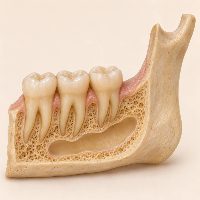
The name refers to its microscopic lining. This distinguishes it from other lesions, including an inflammatory radicular cyst.
Its position, size and nearby structures guide investigation. Examination of a tissue sample establishes the diagnosis.
Surgical management is tailored to the individual. Longer-term checks remain important after treatment because recurrence is possible.
The name refers to its microscopic lining. This distinguishes it from other lesions, including an inflammatory radicular cyst.
Its position, size and nearby structures guide investigation. Examination of a tissue sample establishes the diagnosis.
Surgical management is tailored to the individual. Longer-term checks remain important after treatment because recurrence is possible.
Assessment of findings, images and tissue together.
No. Agreed checks still matter when there are no symptoms.
The responsible dental or surgical team.
Three steps. No audio.
Each scene can also be selected manually.

Silent educational film with German on-screen text and simplified teaching images. Not a patient case or a promised treatment outcome. The full English explanation is available as text.
A radicular cyst is linked to inflammation associated with a tooth whose pulp has died. A keratocyst is a different cyst type with its own tissue features and follow-up considerations.
No. The term names a type of cyst. It can nevertheless grow significantly within the jaw and requires professional assessment.
No. Different jaw lesions may look similar. Microscopic tissue examination is central to the precise diagnosis.
The treating team sets the schedule according to the findings, treatment and progress. Having no symptoms does not replace those checks.
You can bring this question to your appointment:
What tissue diagnosis was established, and which follow-up plan fits this finding?
An individual examination can clarify the findings and suitable next steps.
Arrange an assessmentAppointment selection at DoctolibA radicular cyst is linked to inflammation associated with a tooth whose pulp has died. A keratocyst is a different cyst type with its own tissue features and follow-up considerations.
Editorial research: . General information, not an individual diagnosis or treatment recommendation. Teaching illustrations are simplified and do not depict a patient case.
If you turn this on, this website stores which guides you open, separately from advertising preferences. The markers expire after a fixed 30-day period from activation. The list is not sent to us by this feature. You can turn it off here and delete the markers.
Other people using the same browser profile can also see the markers. German and English versions share one marker. Expired entries are removed the next time you open a page with this feature; deleting browser data may remove them earlier. “Viewed” means opened, not read or completed.