Is there a fixed age?
Timing depends on dental development and the individual cleft.

Alveolar cleft bone grafting fills a congenital gap in the tooth-bearing upper jaw with bone. The aim is to create a bony bridge that supports adjacent teeth and can allow a permanent tooth to erupt in the cleft region.
For training & practice ↓
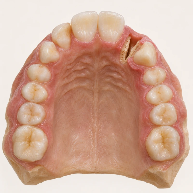
An alveolar cleft interrupts the tooth-bearing upper jaw at one site. Earlier lip or palate repair does not necessarily close that bony gap.
The team often plans grafting before the nearby permanent canine erupts. Tooth position and stage of development guide timing.
During mixed dentition the cleft surgery and orthodontic teams coordinate preparation, grafting and later tooth movement. The outcome is then monitored.
An alveolar cleft interrupts the tooth-bearing upper jaw at one site. Earlier lip or palate repair does not necessarily close that bony gap.
The team often plans grafting before the nearby permanent canine erupts. Tooth position and stage of development guide timing.
During mixed dentition the cleft surgery and orthodontic teams coordinate preparation, grafting and later tooth movement. The outcome is then monitored.
Timing depends on dental development and the individual cleft.
The operation targets the bone of the tooth-bearing ridge.
Its progress is monitored. Orthodontic help may be needed.
Three steps. No audio.
Each scene can also be selected manually.

Silent film with English on-screen text and simplified teaching images.
Alveolar cleft grafting addresses a congenital interruption in the growing dental arch.
The team often uses the child's own bone, for example from the hip. The donor site and possible discomfort are discussed before surgery.
Depending on the position of the jaw segments and teeth, orthodontic preparation may assist treatment.
The centre follows wound healing, bone continuity and subsequent tooth eruption as appropriate.
Ask the cleft centre how orthodontic preparation, surgery and follow-up fit together.
How far has the permanent tooth next to the cleft developed and when is the next review?
Your multidisciplinary cleft team plans individual treatment and follow-up.
Choose an appointmentAppointment selection at DoctolibDental development can be more useful for timing than chronological age alone.
Editorial research: . General information, not an individual diagnosis or treatment recommendation. Teaching illustrations are simplified and do not depict a patient case.
If you turn this on, this website stores which guides you open, separately from advertising preferences. The markers expire after a fixed 30-day period from activation. The list is not sent to us by this feature. You can turn it off here and delete the markers.
Other people using the same browser profile can also see the markers. German and English versions share one marker. Expired entries are removed the next time you open a page with this feature; deleting browser data may remove them earlier. “Viewed” means opened, not read or completed.