Who may find this relevant?
Anyone wanting to understand the inner surfaces of the mouth or a change there.

Oral mucosa is the moist tissue lining many internal mouth surfaces, including the cheeks, lips and floor of the mouth. The gums are also part of it. It protects underlying tissues; its appearance and mobility vary by location.
For training & practice ↓
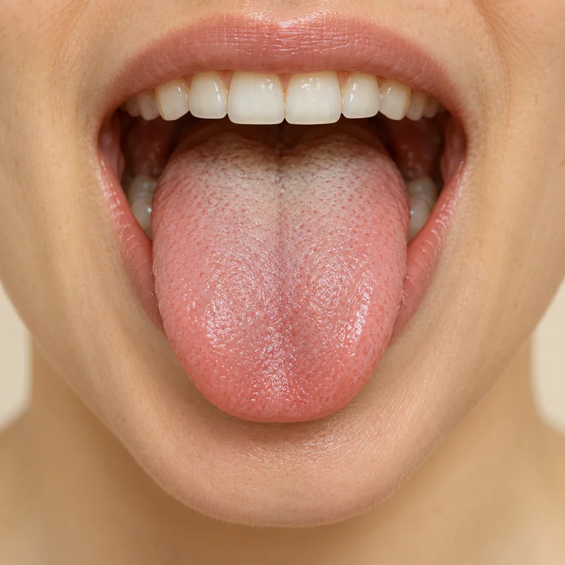
The inside of the lips and cheeks, floor of the mouth, palate and tongue are different regions. Their lining does not look identical everywhere.
The outer cell layer forms a barrier. Moisture and healthy tissue help accommodate friction during eating and speaking. Dry or irritated areas may become uncomfortable.
A sore, lump or red or white patch lasting about two weeks should be assessed. Seek earlier care for rapid worsening, substantial pain or bleeding; breathing difficulty is an emergency.
Anyone wanting to understand the inner surfaces of the mouth or a change there.
Mention the location, onset, changes, symptoms, medicines and possible sources of irritation.
For unexplained changes or an area lasting about two weeks; sooner with significant symptoms.
Three steps. No audio.
Each scene can also be selected manually.

Silent educational film with German on-screen text and simplified teaching images. Not a patient case or a promised treatment outcome. The full English explanation is available below.
The inside of the lips and cheeks, floor of the mouth, palate and tongue are different regions. Their lining does not look identical everywhere.
The outer cell layer forms a barrier. Moisture and healthy tissue help accommodate friction during eating and speaking. Dry or irritated areas may become uncomfortable.
A sore, lump or red or white patch lasting about two weeks should be assessed. Seek earlier care for rapid worsening, substantial pain or bleeding; breathing difficulty is an emergency.
A coating sits on a surface; a mucosal change involves the tissue itself. Do not test removability by forceful scraping.
Gums are one part of it. Oral mucosa includes other surfaces, including areas without teeth.
No. Irritation, infections and other changes may look similar. Persistent or unexplained findings still need examination.
No. Do not scrape, squeeze or apply irritating home remedies. Describe the area and how long it has been present at the appointment.
An editorially suggested question for a discussion with Dr med dent Niusha Barahmand Pour:
What change is present here, and would a review or further investigation be appropriate?
An area is not healing or feels different? Have it examined rather than choosing a diagnosis from pictures.
Arrange an oral examinationAppointment selection at DoctolibA coating sits on a surface; a mucosal change involves the tissue itself. Do not test removability by forceful scraping.
Editorial research: . General information, not an individual diagnosis or treatment recommendation. Teaching illustrations are simplified and do not depict a patient case.