What defines breakdown?
Previously present surface structure is lost after eruption.

Post-eruptive enamel breakdown is loss of enamel after a tooth has emerged. In hypomineralisation, enamel that was initially present but more porous can break down under chewing forces.
For training & practice ↓
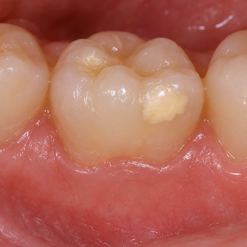
A white to yellow-brown area may indicate altered mineralisation. Colour alone does not constitute breakdown while the surface remains intact.
Some weaker enamel may be lost from a chewing surface. More yellowish dentin may become visible below and the tooth may become sensitive.
The team assesses extent, pain, decay and loading. Depending on the findings, preventive protection or restorative care may be appropriate.
A white to yellow-brown area may indicate altered mineralisation. Colour alone does not constitute breakdown while the surface remains intact.
Some weaker enamel may be lost from a chewing surface. More yellowish dentin may become visible below and the tooth may become sensitive.
The team assesses extent, pain, decay and loading. Depending on the findings, preventive protection or restorative care may be appropriate.
Previously present surface structure is lost after eruption.
Yes. Examination assesses breakdown and possible decay separately.
It may matter alongside surface integrity, symptoms and loading.
Three steps. No audio.
Each scene can also be selected manually.

Silent film with English on-screen text and simplified teaching images.
Too little enamel formed during development. In post-eruptive breakdown, enamel that was present is lost later.
No. Colour and surface loss are separate findings. The team checks whether enamel has actually been lost.
Hypomineralised enamel can break down without preceding decay. Additional decay can develop later at a site that is difficult to clean.
In a follow-up study of children with MIH, yellow opacities more often deteriorated than white ones. Colour can inform review but cannot predict the future of a single tooth.
A question to bring along
How can this sensitive area be protected and when should it be reviewed?
Crumbling enamel or pain during brushing warrants early assessment.
Choose an appointmentAppointment selection at DoctolibColour, timing and surface integrity are recorded separately. A single photograph often cannot establish the diagnosis.
Editorial research: . General information, not an individual diagnosis or treatment recommendation. Teaching illustrations are simplified and do not depict a patient case.
If you turn this on, this website stores which guides you open, separately from advertising preferences. The markers expire after a fixed 30-day period from activation. The list is not sent to us by this feature. You can turn it off here and delete the markers.
Other people using the same browser profile can also see the markers. German and English versions share one marker. Expired entries are removed the next time you open a page with this feature; deleting browser data may remove them earlier. “Viewed” means opened, not read or completed.