Always visible?
No.

Hidden caries is a descriptive term for lesions that are difficult to notice at a glance. Assessment can be challenging between teeth or beneath apparently intact enamel. An unremarkable appearance alone cannot rule decay out.
For training & practice ↓
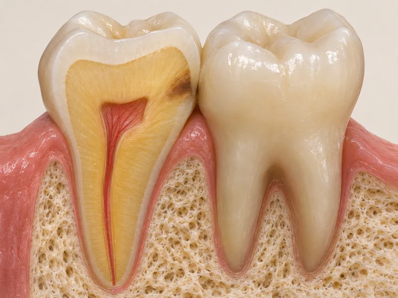
The visible surface does not always reveal the full extent inside a tooth.
Cleaning, drying and careful examination help assessment. Radiographs are added only when appropriately indicated.
Site, depth and activity are considered together. A dark area on a photograph or radiograph is not automatically decay.
No.
No.
Only after individual justification.
Three steps. No audio.
Each scene can also be selected manually.

Silent educational film with German on-screen text and simplified teaching images. Not a patient case or a promised treatment outcome. The full English explanation is available below.
The visible surface does not always reveal the full extent inside a tooth.
Cleaning, drying and careful examination help assessment. Radiographs are added only when appropriately indicated.
Site, depth and activity are considered together. A dark area on a photograph or radiograph is not automatically decay.
A finding may exist without being directly visible. Conversely, a dark image area does not prove disease.
No. Findings, risk and existing images guide the decision.
Yes. Lack of pain does not rule out a carious lesion.
No. A photograph cannot replace examination and any necessary additional tests.
Editorially suggested question to discuss with Dr Niusha Barahmand Pour:
Which examination is appropriate for this area, and why?
We can assess a concerning area and explain which additional tests are useful.
Have suspected decay assessedAppointment selection at DoctolibA finding may exist without being directly visible. Conversely, a dark image area does not prove disease.
Editorial research: . General information, not an individual diagnosis or treatment recommendation. Teaching illustrations are simplified and do not depict a patient case.