What is the aim?
To assess and, where appropriate, treat selected internal joint problems.

TMJ arthroscopy introduces a very small optical instrument into the jaw joint through an access point in front of the ear. It provides a view inside the joint and may be combined with treatments such as lavage or release of adhesions. It is considered for selected joint disorders. It is not a routine investigation for every click and does not replace careful assessment.
For training & practice ↓
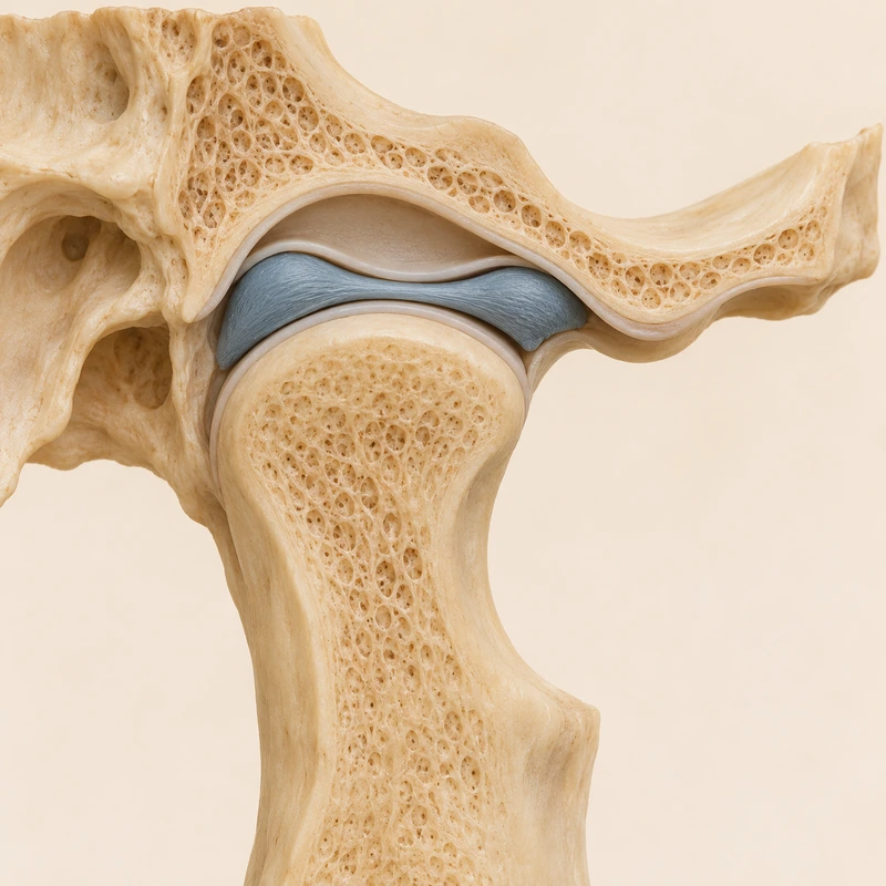
The decision depends on symptoms, examination and imaging where appropriate. Benefits, alternatives and risks are discussed before surgery. Not every altered disc seen on an image causes symptoms that need treatment.
A small camera transmits images from the joint space. Depending on findings, fine instruments and lavage may also be used. The treating team plans the scope and anaesthesia; not every problem can be resolved arthroscopically.
Discomfort and swelling can occur afterwards. Rare injury to nerves or ear structures is possible, occasionally with lasting effects. Follow-up and suitable movement advice matter; a pain-free result is not guaranteed.
The decision depends on symptoms, examination and imaging where appropriate. Benefits, alternatives and risks are discussed before surgery. Not every altered disc seen on an image causes symptoms that need treatment.
A small camera transmits images from the joint space. Depending on findings, fine instruments and lavage may also be used. The treating team plans the scope and anaesthesia; not every problem can be resolved arthroscopically.
Discomfort and swelling can occur afterwards. Rare injury to nerves or ear structures is possible, occasionally with lasting effects. Follow-up and suitable movement advice matter; a pain-free result is not guaranteed.
To assess and, where appropriate, treat selected internal joint problems.
No. Small access points do not remove the risks of surgery.
No; symptoms and relevant findings determine suitability.
Three steps. No audio.
Each scene can also be selected manually.

Silent educational film with German on-screen text and simplified teaching images. Not a patient case or a promised treatment outcome. The full English explanation is available as text.
Arthroscopy provides a direct view using an optical instrument. Arthrocentesis means joint lavage without necessarily using a camera. They may be combined but are distinct concepts.
MRI provides images without inserting an optical instrument into the joint. Arthroscopy is surgery that permits direct inspection and possible treatment. It is not selected simply because more detailed images are wanted.
No. Arthroscopy uses small access points. Open surgery is a different approach that may be needed for some problems; it is not an automatic part of every arthroscopy.
A team with relevant expertise assesses whether symptoms and findings justify the procedure. Initial assessment may lead to referral. This page does not promise that SMD performs arthroscopy.
You can bring this question to your appointment:
What additional information or treatment would arthroscopy offer, and what alternatives and risks should we consider?
An individual assessment can clarify the findings and whether referral is needed.
Arrange an assessmentAppointment selection at DoctolibArthroscopy provides a direct view using an optical instrument. Arthrocentesis means joint lavage without necessarily using a camera. They may be combined but are distinct concepts.
Editorial research: . General information, not an individual diagnosis or treatment recommendation. Teaching illustrations are simplified and do not depict a patient case.
If you turn this on, this website stores which guides you open, separately from advertising preferences. The markers expire after a fixed 30-day period from activation. The list is not sent to us by this feature. You can turn it off here and delete the markers.
Other people using the same browser profile can also see the markers. German and English versions share one marker. Expired entries are removed the next time you open a page with this feature; deleting browser data may remove them earlier. “Viewed” means opened, not read or completed.