How is the image captured?
For example, by a sensor or phosphor plate.

Digital radiography records and stores an X-ray image as digital data. Dental systems include electronic sensors and storage phosphor plates that are scanned after exposure. The examination still uses X-rays: digital refers to image acquisition and processing. It facilitates storage and comparison but does not replace a justified indication or professional interpretation.
For training & practice ↓
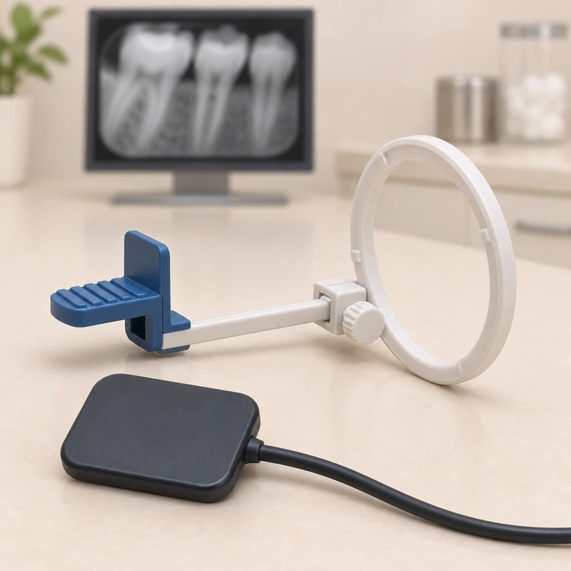
An electronic sensor supplies image data directly. A phosphor plate first stores the exposure pattern and is then scanned. Both routes produce a digital radiographic image.
Brightness, contrast and magnification can help viewing. They cannot create information that was never captured. Original data and adequate image quality remain essential.
Digital technology can reduce exposure but does not eliminate radiation. The clinical question, suitable settings and available previous images determine whether and how to take an X-ray.
An electronic sensor supplies image data directly. A phosphor plate first stores the exposure pattern and is then scanned. Both routes produce a digital radiographic image.
Brightness, contrast and magnification can help viewing. They cannot create information that was never captured. Original data and adequate image quality remain essential.
Digital technology can reduce exposure but does not eliminate radiation. The clinical question, suitable settings and available previous images determine whether and how to take an X-ray.
For example, by a sensor or phosphor plate.
No; it remains an X-ray examination.
Images can be stored, compared and shared.
Three steps. No audio.
Each scene can also be selected manually.

Silent educational film with German on-screen text and simplified teaching images. Not a patient case or a promised treatment outcome. The full English explanation is available as text.
Digital describes the data format. An X-ray sensor detects ionising radiation. Optical methods such as photographs and intraoral scans use a different principle.
No. A single two-dimensional dental radiograph can be digital. CBCT is a three-dimensional acquisition method requiring its own justification.
Not automatically. Receptor type, settings and the diagnostic task matter. Increasing exposure simply to make an image look less noisy should be avoided.
Digital data make transfer easier. Ask which original images and information are needed for further care; sharing suitable previous images may avoid unnecessary repeats.
You can bring this question to your appointment:
Are suitable previous images available, and which digital image would answer the current question?
An individual assessment can clarify the findings and whether referral is needed.
Arrange an assessmentAppointment selection at DoctolibDigital describes the data format. An X-ray sensor detects ionising radiation. Optical methods such as photographs and intraoral scans use a different principle.
Editorial research: . General information, not an individual diagnosis or treatment recommendation. Teaching illustrations are simplified and do not depict a patient case.
If you turn this on, this website stores which guides you open, separately from advertising preferences. The markers expire after a fixed 30-day period from activation. The list is not sent to us by this feature. You can turn it off here and delete the markers.
Other people using the same browser profile can also see the markers. German and English versions share one marker. Expired entries are removed the next time you open a page with this feature; deleting browser data may remove them earlier. “Viewed” means opened, not read or completed.