2D or 3D?
A two-dimensional side view.

A lateral cephalometric radiograph is a standardised side-view X-ray of the head. It can show relationships between jaws, teeth and skull structures for selected orthodontic planning questions. It is not a three-dimensional scan.
For training & practice ↓
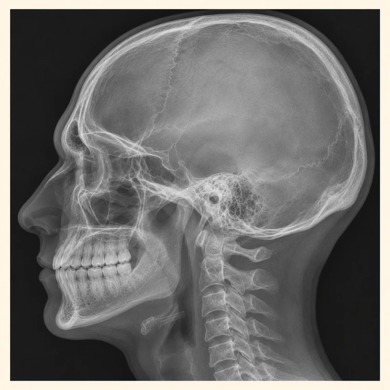
Structures are represented in two dimensions. The right and left sides may overlap. Accurate head positioning helps make images comparable.
Reference points and angles help assess tooth inclinations and jaw relationships. One measurement does not replace examination or assessment of the entire face and bite.
Imaging is selected for a particular question, taking existing images into account. A single cephalogram cannot reliably predict future facial growth.
Structures are represented in two dimensions. The right and left sides may overlap. Accurate head positioning helps make images comparable.
Reference points and angles help assess tooth inclinations and jaw relationships. One measurement does not replace examination or assessment of the entire face and bite.
Imaging is selected for a particular question, taking existing images into account. A single cephalogram cannot reliably predict future facial growth.
A two-dimensional side view.
No.
Tell the team about relevant existing images.
Three steps. No audio.
Each scene can also be selected manually.

Silent educational film with German on-screen text and simplified teaching images. Not a patient case or a promised treatment outcome. The full English explanation is available as text.
A cephalogram is a two-dimensional side projection. CBCT produces a three-dimensional dataset and requires its own justified indication.
No. Need depends on the intended changes and clinical findings, not simply on the fact that braces are planned.
No. A panoramic image provides a different overview of teeth and jaws. A cephalogram shows the head and jaw relationship from the side.
No. Individual images and measurements do not provide a reliable personal growth prediction.
You can bring this question to your appointment:
Which planning question would this image answer that other findings cannot adequately resolve?
Discuss your individual findings and whether a referral is appropriate.
Discuss your findingsAppointment selection at DoctolibA cephalogram is a two-dimensional side projection. CBCT produces a three-dimensional dataset and requires its own justified indication.
Editorial research: . General information, not an individual diagnosis or treatment recommendation. Teaching illustrations are simplified and do not depict a patient case.