Can the colour be natural?
Pigmentation can be a normal variation.

Melanin can give healthy gums patches of light to dark brown. A new, isolated mark may have a different cause. Its distribution, history and an examination of the mouth help identify it.
For training & practice ↓
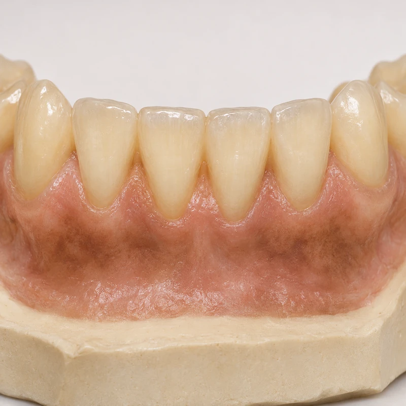
Melanin may produce brown bands or areas of varying size on both sides of the gums.
The dentist checks whether the colour is widespread or confined to one spot.
Tell the dentist when you first noticed the patch and whether it has grown. Earlier photographs may help show changes.
Melanin may produce brown bands or areas of varying size on both sides of the gums.
The dentist checks whether the colour is widespread or confined to one spot.
Tell the dentist when you first noticed the patch and whether it has grown. Earlier photographs may help show changes.
Pigmentation can be a normal variation.
Foreign material and blood can also look dark.
Arrange an examination, especially if its shape or size changes.
Three steps. No audio.
Each scene can also be selected manually.

Silent film with English on-screen text and simplified teaching images.
Pigmentation describes a deposit of colour. Swelling, pain or bleeding are assessed separately.
A photo can document change. Diagnosis may require an examination and, if the finding remains unclear, a tissue sample.
Melanin lies in the tissue. Brushing cannot remove it.
They describe diagnoses in sampled lesions. Unusual patches are more likely to be biopsied than ordinary physiological pigmentation.
A question to bring along
What would make you recommend a biopsy of this patch?
Arrange an oral examination for a new or changing mark.
Choose an appointmentAppointment selection at DoctolibAppearance narrows the possibilities. History and, when needed, tissue examination help distinguish uncertain causes.
Editorial research: . General information, not an individual diagnosis or treatment recommendation. Teaching illustrations are simplified and do not depict a patient case.
If you turn this on, this website stores which guides you open, separately from advertising preferences. The markers expire after a fixed 30-day period from activation. The list is not sent to us by this feature. You can turn it off here and delete the markers.
Other people using the same browser profile can also see the markers. German and English versions share one marker. Expired entries are removed the next time you open a page with this feature; deleting browser data may remove them earlier. “Viewed” means opened, not read or completed.