Where is the upper end?
At the nasal floor, with a variable number of openings.

The nasopalatine canal is a bony passage behind the upper central incisors. It runs between the floor of the nose and the hard palate and carries nerves and vessels. It may divide towards the nose.
For training & practice ↓
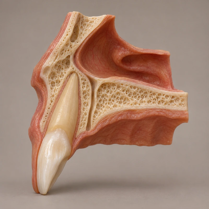
The canal reaches the nasal floor above. Three-dimensional imaging studies show that the number of openings there varies.
On the oral side the canal opens just behind the upper central incisors. This palatal opening is the incisive foramen.
Shape and direction vary between people. Planning a procedure in the anterior maxilla therefore depends on its relationship to the proposed site.
The canal reaches the nasal floor above. Three-dimensional imaging studies show that the number of openings there varies.
On the oral side the canal opens just behind the upper central incisors. This palatal opening is the incisive foramen.
Shape and direction vary between people. Planning a procedure in the anterior maxilla therefore depends on its relationship to the proposed site.
At the nasal floor, with a variable number of openings.
The incisive foramen.
The shape, direction and dimensions of the canal.
Three steps. No audio.
Each scene can also be selected manually.

Silent educational film with German on-screen text and simplified teaching images. Not a patient case or a promised treatment outcome. The full English explanation is available as text.
The canal is normal anatomy. A cyst in this region is a separate finding requiring its own assessment.
The canal reaches the nasal floor above. Three-dimensional imaging studies show that the number of openings there varies.
On the oral side the canal opens just behind the upper central incisors. This palatal opening is the incisive foramen.
Shape and direction vary between people. Planning a procedure in the anterior maxilla therefore depends on its relationship to the proposed site.
It runs within the maxillary bone behind the teeth. Each incisor has its own internal canal system.
The canal is normal anatomy. A cyst in this region is a separate finding requiring its own assessment.
A question you could ask
How does the canal relate to the planned procedure in the front of my upper jaw?
When planning care near the upper incisors, ask to see the canal in relation to the proposed site.
Discuss at a dental appointmentAppointment selection at DoctolibThe canal is normal anatomy. A cyst in this region is a separate finding requiring its own assessment.
Editorial research: . General information, not an individual diagnosis or treatment recommendation. Teaching illustrations are simplified and do not depict a patient case.
If you turn this on, this website stores which guides you open, separately from advertising preferences. The markers expire after a fixed 30-day period from activation. The list is not sent to us by this feature. You can turn it off here and delete the markers.
Other people using the same browser profile can also see the markers. German and English versions share one marker. Expired entries are removed the next time you open a page with this feature; deleting browser data may remove them earlier. “Viewed” means opened, not read or completed.