Where does it occur?
Around the incisive canal in the anterior upper jaw.

A nasopalatine duct cyst is a developmental cyst that does not originate from a tooth. It occurs around the incisive canal in the front of the upper jaw. Because it can lie close to the upper incisor roots, it may resemble inflammation arising from those teeth. The teeth and the lesion therefore need separate assessment.
For training & practice ↓
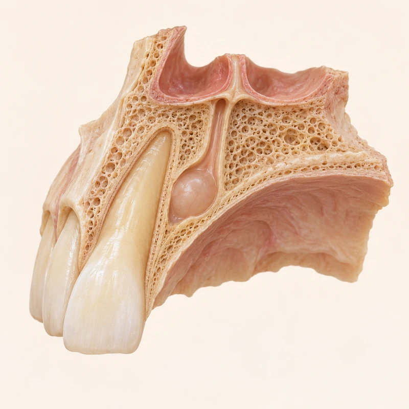
The incisive canal lies in the midline of the anterior upper jaw. A cyst may develop from embryonic tissue remnants in this region.
Pulp sensibility testing and appropriate radiographs help assess whether a tooth is involved. Proximity to roots does not establish a dental cause.
Tissue examination complements the clinical and imaging findings. Any surgical approach is planned for the individual situation.
The incisive canal lies in the midline of the anterior upper jaw. A cyst may develop from embryonic tissue remnants in this region.
Pulp sensibility testing and appropriate radiographs help assess whether a tooth is involved. Proximity to roots does not establish a dental cause.
Tissue examination complements the clinical and imaging findings. Any surgical approach is planned for the individual situation.
Around the incisive canal in the anterior upper jaw.
Not for this cyst type; any accompanying tooth disease is assessed separately.
Combining tooth tests, imaging and tissue findings.
Three steps. No audio.
Each scene can also be selected manually.

Silent educational film with German on-screen text and simplified teaching images. Not a patient case or a promised treatment outcome. The full English explanation is available as text.
The nasopalatine duct cyst is a nonodontogenic developmental cyst. A radicular cyst is an inflammatory lesion associated with a tooth whose pulp has died.
Not automatically. This cyst does not arise from the dental pulp. Root canal treatment requires a separate tooth-related indication.
No. It may be found incidentally, although swelling or pain can occur. Symptoms alone do not identify the cyst type.
No. Overlapping structures influence the shape seen. Clinical assessment and tissue diagnosis remain necessary.
You can bring this question to your appointment:
Is the lesion independent of the incisor, and which findings establish that distinction?
An individual examination can clarify the findings and suitable next steps.
Arrange an assessmentAppointment selection at DoctolibThe nasopalatine duct cyst is a nonodontogenic developmental cyst. A radicular cyst is an inflammatory lesion associated with a tooth whose pulp has died.
Editorial research: . General information, not an individual diagnosis or treatment recommendation. Teaching illustrations are simplified and do not depict a patient case.
If you turn this on, this website stores which guides you open, separately from advertising preferences. The markers expire after a fixed 30-day period from activation. The list is not sent to us by this feature. You can turn it off here and delete the markers.
Other people using the same browser profile can also see the markers. German and English versions share one marker. Expired entries are removed the next time you open a page with this feature; deleting browser data may remove them earlier. “Viewed” means opened, not read or completed.