What is a macule?
A flat patch of changed colour.

An oral melanotic macule is a localised flat brown-to-dark patch of oral mucosa caused by increased deposition of the body’s own melanin. A confirmed macule is benign and usually symptom-free. Nevertheless, a new or changing pigmented patch must not be self-diagnosed by colour: other causes, including rare malignant lesions, need to be excluded.
For training & practice ↓
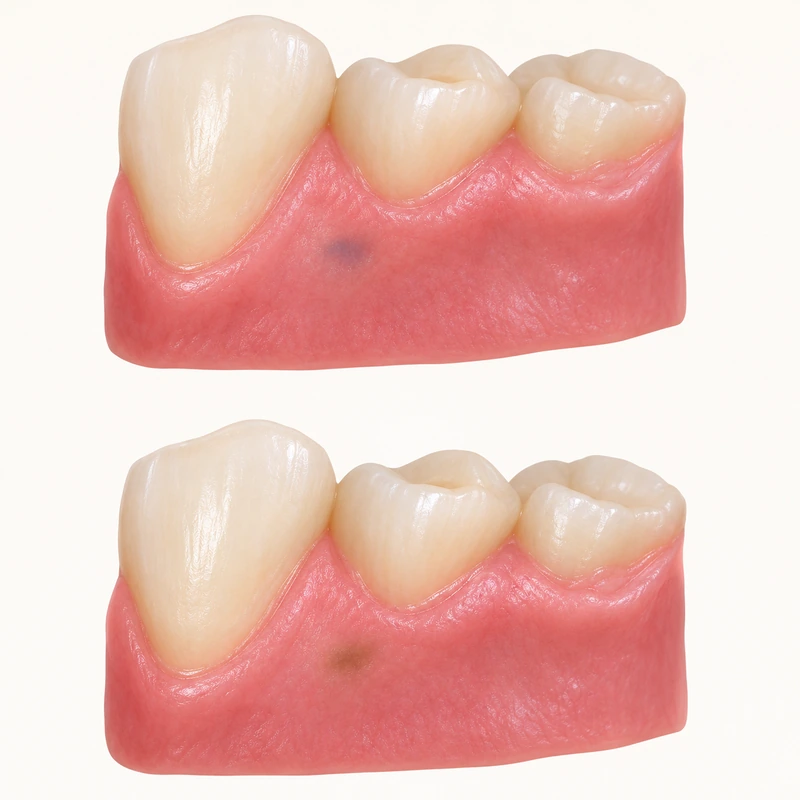
Melanin is a pigment produced by the body. A melanotic macule has locally increased pigmentation. Similar patches may occur on the lip or within the mouth.
Assessment considers location, size, borders and timing. A new or enlarging patch, or a change in colour or shape, warrants focused professional examination.
Uncertain or suspicious pigmentation may need a biopsy. A confirmed benign macule can be monitored. Home bleaching or caustic removal is not suitable treatment.
Melanin is a pigment produced by the body. A melanotic macule has locally increased pigmentation. Similar patches may occur on the lip or within the mouth.
Assessment considers location, size, borders and timing. A new or enlarging patch, or a change in colour or shape, warrants focused professional examination.
Uncertain or suspicious pigmentation may need a biopsy. A confirmed benign macule can be monitored. Home bleaching or caustic removal is not suitable treatment.
A flat patch of changed colour.
The body’s own melanin.
Seek examination; do not bleach it yourself.
Three steps. No audio.
Each scene can also be selected manually.

Silent educational film with German on-screen text and simplified teaching images. Not a patient case or a promised treatment outcome. The full English explanation is available as text.
A macule is a flat colour change, not a palpable lump. This description does not establish the cause. Distinguishing a pigmented naevus or another lesion may require further investigation.
No. An amalgam tattoo contains embedded foreign material; a melanotic macule involves the body’s own melanin. Similar colours do not allow reliable differentiation.
That depends on location, history and diagnostic certainty. Biopsy can be important for suspicious or changing lesions. The examining team makes that decision, not an online colour comparison.
No. Absence of pain does not reliably identify the cause. An unfamiliar or changing patch needs examination even if it causes no discomfort.
No. Melanotic macules also occur in areas without sun exposure. A possible role for sunlight on the outer lip should not be assumed for the oral mucosa.
You can bring this question to your appointment:
Can this pigmentation be identified confidently, and do we need a biopsy or follow-up?
An individual examination can clarify the findings and suitable next steps.
Arrange an assessmentAppointment selection at DoctolibA macule is a flat colour change, not a palpable lump. This description does not establish the cause. Distinguishing a pigmented naevus or another lesion may require further investigation.
Editorial research: . General information, not an individual diagnosis or treatment recommendation. Teaching illustrations are simplified and do not depict a patient case.
If you turn this on, this website stores which guides you open, separately from advertising preferences. The markers expire after a fixed 30-day period from activation. The list is not sent to us by this feature. You can turn it off here and delete the markers.
Other people using the same browser profile can also see the markers. German and English versions share one marker. Expired entries are removed the next time you open a page with this feature; deleting browser data may remove them earlier. “Viewed” means opened, not read or completed.