Which projection is in front?
The coronoid process.

The coronoid process is the front projection at the top of the mandibular ramus. It provides an attachment for the temporalis muscle, which transmits force to the mandible when closing the jaw.
For training & practice ↓
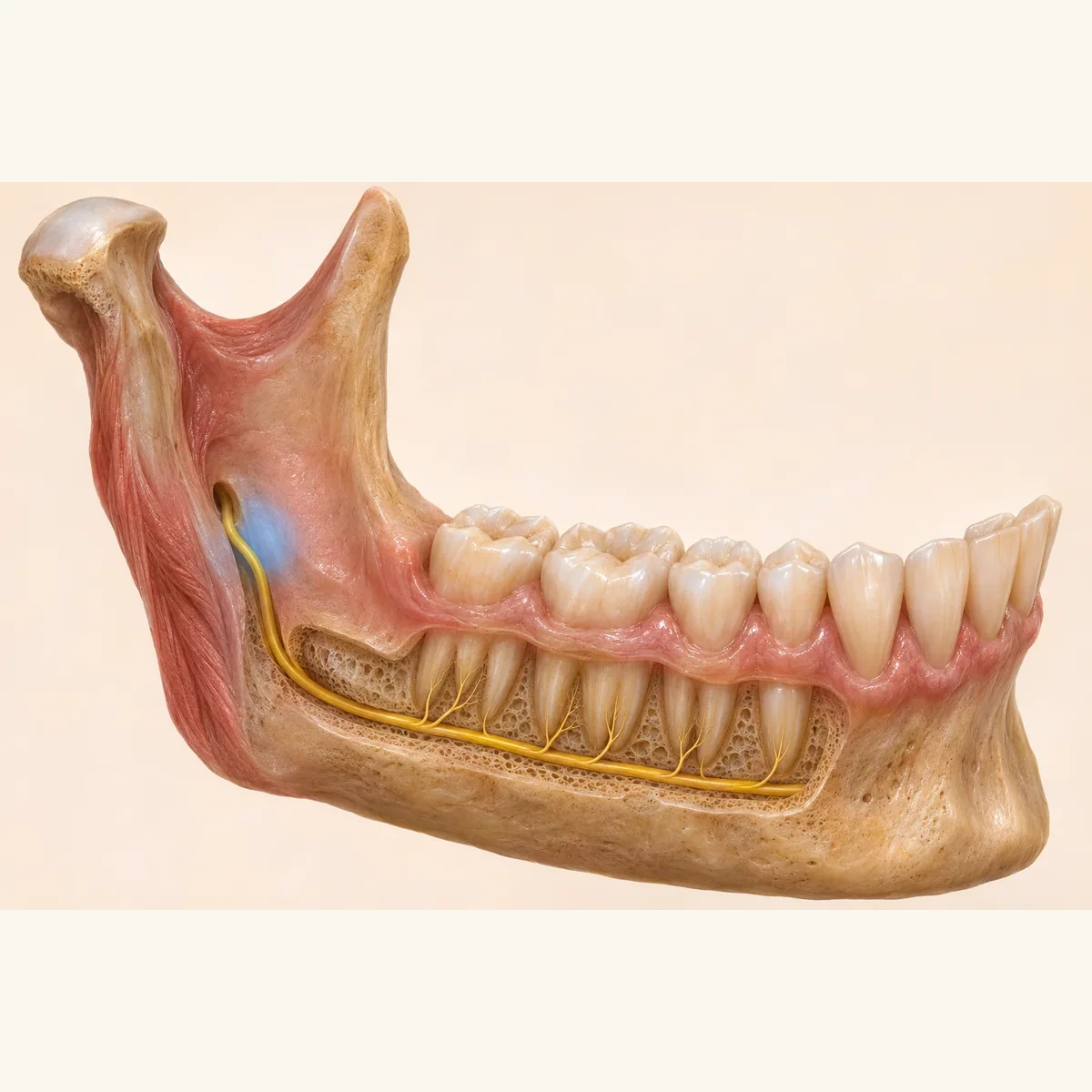
Two projections rise from the ramus. In side view, the flatter anterior coronoid process can be distinguished from the joint process behind it.
The temporalis tendon reaches the coronoid process. The muscle works with other chewing muscles to elevate the jaw.
The mandibular notch separates the coronoid and condylar processes and helps identify them on a bone model.
Two projections rise from the ramus. In side view, the flatter anterior coronoid process can be distinguished from the joint process behind it.
The temporalis tendon reaches the coronoid process. The muscle works with other chewing muscles to elevate the jaw.
The mandibular notch separates the coronoid and condylar processes and helps identify them on a bone model.
The coronoid process.
The temporalis muscle.
The mandibular notch, an indentation in the bone.
Three steps. No audio.
Each scene can also be selected manually.

Silent educational film with German on-screen text and simplified teaching images. Not a patient case or a promised treatment outcome. The full English explanation is available as text.
The joint head sits on the rear projection. The front projection provides a muscle attachment.
Two projections rise from the ramus. In side view, the flatter anterior coronoid process can be distinguished from the joint process behind it.
The temporalis tendon reaches the coronoid process. The muscle works with other chewing muscles to elevate the jaw.
The mandibular notch separates the coronoid and condylar processes and helps identify them on a bone model.
It belongs to the jawbone. A tooth crown is the coronal part of a tooth or a restoration covering it.
The joint head sits on the rear projection. The front projection provides a muscle attachment.
A question you could ask
Which of the two projections is being described on my image?
At an imaging review, ask to see the muscle attachment and the joint head side by side.
Discuss at a dental appointmentAppointment selection at DoctolibThe joint head sits on the rear projection. The front projection provides a muscle attachment.
Editorial research: . General information, not an individual diagnosis or treatment recommendation. Teaching illustrations are simplified and do not depict a patient case.
If you turn this on, this website stores which guides you open, separately from advertising preferences. The markers expire after a fixed 30-day period from activation. The list is not sent to us by this feature. You can turn it off here and delete the markers.
Other people using the same browser profile can also see the markers. German and English versions share one marker. Expired entries are removed the next time you open a page with this feature; deleting browser data may remove them earlier. “Viewed” means opened, not read or completed.