What else is checked?
Other teeth and the pulp spaces.

Radicular dentine dysplasia, or dentine dysplasia type I, is a rare disorder of dentine formation. Crowns may look unremarkable despite markedly shortened roots and greatly reduced pulp spaces. Reduced root support may become apparent in the permanent dentition.
For training & practice ↓
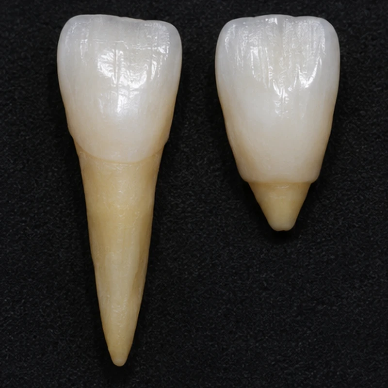
The two models have similarly sized crowns. The short root on the right shows why crown appearance alone tells little about support.
Several roots appear shortened in the simulated radiograph. Type I may show markedly reduced pulp spaces or only narrow remnants.
Stability, surrounding bone, symptoms and signs of inflammation are assessed tooth by tooth. Family history can add useful context.
The two models have similarly sized crowns. The short root on the right shows why crown appearance alone tells little about support.
Several roots appear shortened in the simulated radiograph. Type I may show markedly reduced pulp spaces or only narrow remnants.
Stability, surrounding bone, symptoms and signs of inflammation are assessed tooth by tooth. Family history can add useful context.
Other teeth and the pulp spaces.
A detailed family history.
The outlook for each tooth.
Three steps. No audio.
Each scene can also be selected manually.

Silent film with English on-screen text and simplified teaching images.
Resorption removes previously formed root tissue. Dentine dysplasia affects its development.
No. Developmental stage, other variants and acquired damage can affect shape. The overall pattern matters.
Yes. A multigenerational family study documented autosomal dominant inheritance. An image alone cannot establish inheritance in an individual family.
The diagnosis alone does not decide this. Support, symptoms and treatability guide preservation and monitoring.
A question to bring along
Which of my teeth need closer follow-up?
Earlier radiographs and a family history of similar findings can help assess apparently short roots.
Choose an appointmentAppointment selection at DoctolibRadiographic findings relate to dental hard tissues. They cannot by themselves establish a particular cause of pain or predict when a tooth will be lost.
Editorial research: . General information, not an individual diagnosis or treatment recommendation. Teaching illustrations are simplified and do not depict a patient case.
If you turn this on, this website stores which guides you open, separately from advertising preferences. The markers expire after a fixed 30-day period from activation. The list is not sent to us by this feature. You can turn it off here and delete the markers.
Other people using the same browser profile can also see the markers. German and English versions share one marker. Expired entries are removed the next time you open a page with this feature; deleting browser data may remove them earlier. “Viewed” means opened, not read or completed.