Always after injury?
Injury is one possible association.

Pulp canal obliteration narrows the space inside a tooth through additional hard-tissue formation, sometimes making it barely visible on X-rays. It can follow dental injury. The finding alone means neither a dead pulp nor a need for root canal treatment.
For training & practice ↓
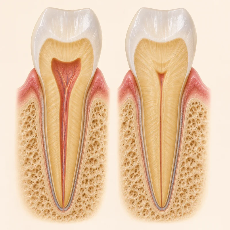
The natural pulp space is affected. This is not an inserted root filling; a remaining canal may not be visible on the X-ray.
The tooth may look more yellow. Colour, symptoms, examination and images are considered together; a single sensibility test may be insufficient.
Without symptoms or evidence of irreversible pulpitis or apical disease, obliteration alone does not indicate treatment. If treatment becomes necessary, locating the narrow canal can be demanding.
The natural pulp space is affected. This is not an inserted root filling; a remaining canal may not be visible on the X-ray.
The tooth may look more yellow. Colour, symptoms, examination and images are considered together; a single sensibility test may be insufficient.
Without symptoms or evidence of irreversible pulpitis or apical disease, obliteration alone does not indicate treatment. If treatment becomes necessary, locating the narrow canal can be demanding.
Injury is one possible association.
No, the tooth’s own hard tissue.
Follow the individually agreed plan.
Three steps. No audio.
Each scene can also be selected manually.

Silent educational film with German on-screen text and simplified teaching images. Not a patient case or a promised treatment outcome. The full English explanation is available as text.
Obliteration describes a narrowing of the internal space; necrosis means tissue death. They can coexist, but are not synonyms.
No. After injury in particular, obliteration may be associated with surviving tissue.
Not solely because of obliteration. Signs of disease and the complete findings guide the decision.
Pain, swelling or a draining sinus need timely assessment. Agreed reviews remain important even without pain.
You can bring this question to your appointment:
Are there signs of disease or is review sufficient, and might specialist treatment be needed?
An examination can clarify the finding and any need for referral.
Arrange an assessmentAppointment selection at DoctolibObliteration describes a narrowing of the internal space; necrosis means tissue death. They can coexist, but are not synonyms.
Editorial research: . General information, not an individual diagnosis or treatment recommendation. Teaching illustrations are simplified and do not depict a patient case.