Radiolucent?
Relatively X-ray-transmitting and usually darker.

Radiolucency describes a relatively X-ray-transmitting structure that appears darker on a conventional radiographic display. Radiopacity means greater X-ray attenuation and a lighter appearance. These terms first describe the image. A dark area is not automatically decay or a cyst: normal anatomy also produces dark and light regions.
For training & practice ↓
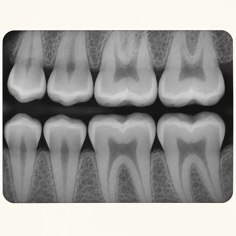
Tissues attenuate X-rays to different degrees. Where more radiation reaches the detector, the usual display is darker. Greater attenuation produces a lighter appearance.
Enamel usually appears lighter than dentine, while the pulp space is darker by comparison. These normal differences provide landmarks before an image is assessed for abnormalities.
Location, shape, borders and surrounding anatomy matter. Symptoms and examination add context. Overlapping structures can also change the appearance of a two-dimensional image.
Tissues attenuate X-rays to different degrees. Where more radiation reaches the detector, the usual display is darker. Greater attenuation produces a lighter appearance.
Enamel usually appears lighter than dentine, while the pulp space is darker by comparison. These normal differences provide landmarks before an image is assessed for abnormalities.
Location, shape, borders and surrounding anatomy matter. Symptoms and examination add context. Overlapping structures can also change the appearance of a two-dimensional image.
Relatively X-ray-transmitting and usually darker.
More X-ray-attenuating and usually lighter.
An image description, not yet a diagnosis.
Three steps. No audio.
Each scene can also be selected manually.

Silent educational film with German on-screen text and simplified teaching images. Not a patient case or a promised treatment outcome. The full English explanation is available as text.
Radiolucent and radiopaque describe appearance relative to surrounding structures. Diagnoses such as decay or a cyst require additional features and clinical context.
No. The normal pulp space, for example, is relatively radiolucent. Darkness alone cannot identify disease.
No. Mineralised tissues such as enamel and bone also attenuate X-rays. Material composition and tissue thickness affect appearance.
A screenshot may alter contrast or crop important details. Assessment requires suitable original images and the clinical examination.
You can bring this question to your appointment:
Which structure are we looking at, and what other findings support your interpretation?
An individual assessment can clarify the findings and whether referral is needed.
Arrange an assessmentAppointment selection at DoctolibRadiolucent and radiopaque describe appearance relative to surrounding structures. Diagnoses such as decay or a cyst require additional features and clinical context.
Editorial research: . General information, not an individual diagnosis or treatment recommendation. Teaching illustrations are simplified and do not depict a patient case.
If you turn this on, this website stores which guides you open, separately from advertising preferences. The markers expire after a fixed 30-day period from activation. The list is not sent to us by this feature. You can turn it off here and delete the markers.
Other people using the same browser profile can also see the markers. German and English versions share one marker. Expired entries are removed the next time you open a page with this feature; deleting browser data may remove them earlier. “Viewed” means opened, not read or completed.