What is insufficient for a diagnosis?
Dark colour alone.

Marginal staining is a discoloured line where a filling meets the tooth. It may be visible while the restoration still functions well. Colour alone does not establish whether decay, a gap or another defect is also present.
For training & practice ↓
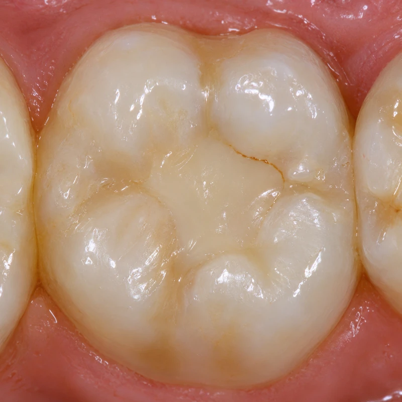
A fine brown line follows part of the filling margin. Its colour is visible here, but the image cannot establish its depth.
The dentist assesses the filling, nearby enamel and surface shape. Further examination may be needed if there is a reason for concern.
An overhang is a physical projection of excess material. A colour difference may also be present at a smooth margin.
A fine brown line follows part of the filling margin. Its colour is visible here, but the image cannot establish its depth.
The dentist assesses the filling, nearby enamel and surface shape. Further examination may be needed if there is a reason for concern.
An overhang is a physical projection of excess material. A colour difference may also be present at a smooth margin.
Dark colour alone.
In the natural grooves of the chewing surface.
Depending on findings, monitoring, polishing or local repair.
Three steps. No audio.
Each scene can also be selected manually.

Silent film with English on-screen text and simplified teaching images.
A gap is a discontinuity at the interface. Staining describes its colour.
The area is cleaned and dried; the dentist assesses continuity, adjacent tooth tissue and change over time. A dark line by itself does not establish decay.
A fissure is a natural groove in the tooth. Marginal staining follows the boundary of a restoration. Either can appear dark and needs assessment in context.
Monitoring or polishing may suit a superficial finding. A localised defect may allow repair. The condition of the whole restoration guides the decision.
A question to bring along
Is the margin only stained, or is there also a contour defect or decay?
If the line is new, is increasing or is accompanied by symptoms, have the tooth checked.
Choose an appointmentAppointment selection at DoctolibFDI criteria evaluate marginal staining and caries at the restoration margin separately. More than one feature can be abnormal.
Editorial research: . General information, not an individual diagnosis or treatment recommendation. Teaching illustrations are simplified and do not depict a patient case.
If you turn this on, this website stores which guides you open, separately from advertising preferences. The markers expire after a fixed 30-day period from activation. The list is not sent to us by this feature. You can turn it off here and delete the markers.
Other people using the same browser profile can also see the markers. German and English versions share one marker. Expired entries are removed the next time you open a page with this feature; deleting browser data may remove them earlier. “Viewed” means opened, not read or completed.