Where?
At the back of the lower jaw.

The retromolar pad is a normal soft-tissue region behind the lower molar area. Mobile mucosa covers loose connective tissue that includes glandular tissue. It provides an anatomical landmark in prosthodontics. In precise terminology it differs from the more anterior, scar-related pear-shaped pad after tooth loss. The name means neither a tumour nor an automatic denture sore.
For training & practice ↓
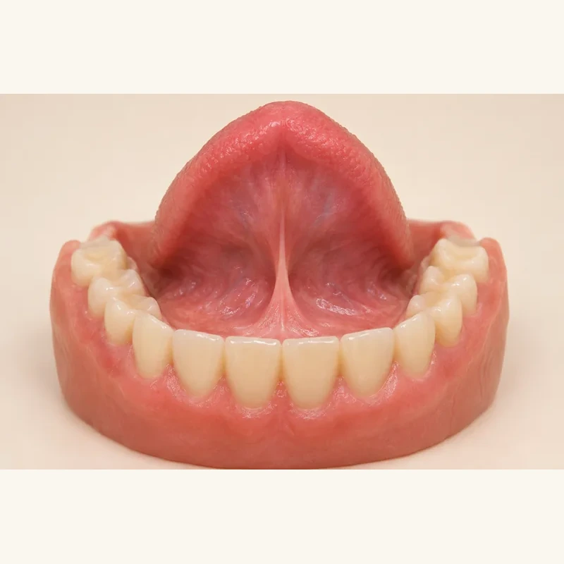
The pad lies at the back of the lower dental arch or edentulous ridge. It is soft tissue, not an additional tooth.
The more mobile retromolar pad is not interchangeable with every adjacent ridge region. Scar-related tissue after tooth loss has a separate technical description.
The region helps orient lower-denture planning. Tender or persistently altered tissue needs examination.
The pad lies at the back of the lower dental arch or edentulous ridge. It is soft tissue, not an additional tooth.
The more mobile retromolar pad is not interchangeable with every adjacent ridge region. Scar-related tissue after tooth loss has a separate technical description.
The region helps orient lower-denture planning. Tender or persistently altered tissue needs examination.
At the back of the lower jaw.
Soft tissue.
No.
Three steps. No audio.
Each scene can also be selected manually.

Silent educational film with German on-screen text and simplified teaching images. Not a patient case or a promised treatment outcome. The full English explanation is available as text.
The pad is a normal structure. A denture sore is tissue irritation and can occur in different locations.
No. Retromolar pad is a normal anatomical term. A new swelling must be distinguished from the normal structure.
Here the term refers to the lower jaw. Posterior upper-jaw structures should not be labelled as the same pad.
It is a posterior mandibular landmark. Individual tissue shape and mobility inform denture design; a picture cannot determine the border.
The specialist glossary distinguishes them. The pear-shaped pad is the scar-related posterior ridge area after removal of the last molar. The retromolar pad lies behind it and is more mobile.
New lumps, persistent red or white patches, and ulcers lasting over three weeks require assessment. Suspected denture sores should also be checked instead of attributing every change to pressure.
You can bring this question to your appointment:
Is this normal tissue, a denture sore or a change needing separate assessment?
A dental examination can clarify how this relates to your findings and treatment planning.
Discuss your dental findingsAppointment selection at DoctolibThe pad is a normal structure. A denture sore is tissue irritation and can occur in different locations.
Editorial research: . General information, not an individual diagnosis or treatment recommendation. Teaching illustrations are simplified and do not depict a patient case.
If you turn this on, this website stores which guides you open, separately from advertising preferences. The markers expire after a fixed 30-day period from activation. The list is not sent to us by this feature. You can turn it off here and delete the markers.
Other people using the same browser profile can also see the markers. German and English versions share one marker. Expired entries are removed the next time you open a page with this feature; deleting browser data may remove them earlier. “Viewed” means opened, not read or completed.