Visible without magnification?
No; they are microscopic.

Enamel prisms, also called enamel rods, are microscopic elongated structural units of tooth enamel. They contain tightly packed mineral crystallites, mainly apatite. Crystal orientation differs within neighbouring prisms and the material between them. Enamel is therefore a spatially organised composite rather than a uniform glass-like layer.
For training & practice ↓
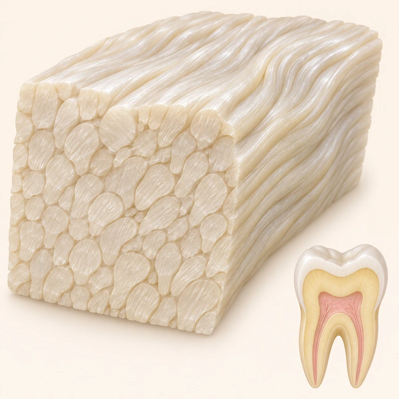
Many small crystallites together form a prism. The teaching model greatly enlarges the microstructure; individual prisms cannot be seen with the naked eye.
Prisms are not aligned identically everywhere. Depending on the site and section, they may look elongated, rounded or keyhole-like. A diagram is only a simplified view.
Prismatic organisation is part of normal enamel. Visible lines, patches or symptoms cannot be diagnosed from a picture of this microstructure alone.
Many small crystallites together form a prism. The teaching model greatly enlarges the microstructure; individual prisms cannot be seen with the naked eye.
Prisms are not aligned identically everywhere. Depending on the site and section, they may look elongated, rounded or keyhole-like. A diagram is only a simplified view.
Prismatic organisation is part of normal enamel. Visible lines, patches or symptoms cannot be diagnosed from a picture of this microstructure alone.
No; they are microscopic.
No; mineralised structure.
No; their course varies.
Three steps. No audio.
Each scene can also be selected manually.

Silent educational film with German on-screen text and simplified teaching images. Not a patient case or a promised treatment outcome. The full English explanation is available as text.
A crystallite is a tiny mineral building block. An enamel prism contains many crystallites arranged together.
No. They are mineralised structural units. The cells that form enamel are called ameloblasts; these are not the same as the finished prisms.
No. Appearance depends on location, course and sectioning, among other factors. Regular patterns in teaching diagrams are simplifications.
Individual prisms are too small for that. Visible surface features are on a different scale and should be assessed clinically if there is concern.
You can bring this question to your appointment:
What visible change is actually present, and how is enamel structure relevant to it?
An individual assessment can clarify the findings and whether referral is needed.
Arrange an assessmentAppointment selection at DoctolibA crystallite is a tiny mineral building block. An enamel prism contains many crystallites arranged together.
Editorial research: . General information, not an individual diagnosis or treatment recommendation. Teaching illustrations are simplified and do not depict a patient case.
If you turn this on, this website stores which guides you open, separately from advertising preferences. The markers expire after a fixed 30-day period from activation. The list is not sent to us by this feature. You can turn it off here and delete the markers.
Other people using the same browser profile can also see the markers. German and English versions share one marker. Expired entries are removed the next time you open a page with this feature; deleting browser data may remove them earlier. “Viewed” means opened, not read or completed.