What is examined?
The salivary gland duct.

Sialendoscopy examines the ducts of major salivary glands using a very thin endoscope introduced through the natural opening inside the mouth. It can investigate and sometimes treat selected obstructions, including certain stones or narrowings. Suitability depends on the findings.
For training & practice ↓
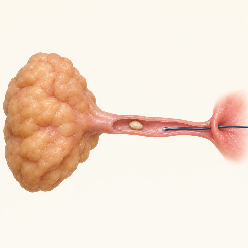
A small optical instrument provides a view inside the duct. Symptoms, examination and appropriate imaging help establish beforehand whether obstruction is present and endoscopy may help.
Selected obstructions can be treated through the endoscope. Not every stone or narrowing is accessible; combined or alternative approaches may be needed.
The aim is often to relieve symptoms while preserving the gland. Swelling, infection, duct injury or later scarring can occur; success and gland preservation are not guaranteed.
A small optical instrument provides a view inside the duct. Symptoms, examination and appropriate imaging help establish beforehand whether obstruction is present and endoscopy may help.
Selected obstructions can be treated through the endoscope. Not every stone or narrowing is accessible; combined or alternative approaches may be needed.
The aim is often to relieve symptoms while preserving the gland. Swelling, infection, duct injury or later scarring can occur; success and gland preservation are not guaranteed.
The salivary gland duct.
Yes, for suitable findings.
No. Limitations and alternatives are discussed beforehand.
Three steps. No audio.
Each scene can also be selected manually.

Silent educational film with German on-screen text and simplified teaching images. Not a patient case or a promised treatment outcome. The full English explanation is available as text.
Ultrasound assesses the gland and surrounding tissues from outside using sound waves. Sialendoscopy looks directly inside the duct and may allow selected treatments. They answer different questions.
No. It works within the duct and often aims to avoid gland removal. Unsuitable findings or incomplete treatment may require other approaches.
No. The cause must first be established. Acute inflammation can make intervention more difficult; the responsible team determines timing and technique.
There is no universal rule. Anaesthesia and the setting depend on the gland, findings, planned intervention and individual circumstances.
You can bring this question to your appointment:
What obstruction is suspected, and what are the benefits and limitations of sialendoscopy for me?
An individual examination can clarify the findings and suitable next steps.
Arrange an assessmentAppointment selection at DoctolibUltrasound assesses the gland and surrounding tissues from outside using sound waves. Sialendoscopy looks directly inside the duct and may allow selected treatments. They answer different questions.
Editorial research: . General information, not an individual diagnosis or treatment recommendation. Teaching illustrations are simplified and do not depict a patient case.
If you turn this on, this website stores which guides you open, separately from advertising preferences. The markers expire after a fixed 30-day period from activation. The list is not sent to us by this feature. You can turn it off here and delete the markers.
Other people using the same browser profile can also see the markers. German and English versions share one marker. Expired entries are removed the next time you open a page with this feature; deleting browser data may remove them earlier. “Viewed” means opened, not read or completed.