Where are voxels used?
For example, in CBCT and CT datasets.

A voxel is a small volume element in a three-dimensional image dataset. In cone-beam CT, the examined space is divided computationally into these elements, each assigned a grey value. Unlike a pixel, a voxel also has depth. Voxel size influences potential detail but is not the same as the image’s actual spatial resolution.
For training & practice ↓
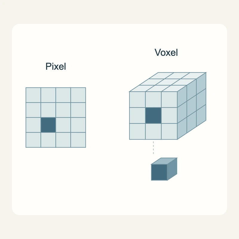
A pixel belongs to a flat image grid; a voxel belongs to a volume grid. Many volume elements together make up the reconstructed CBCT dataset.
Small voxels can support fine detail. Motion, image noise and other equipment properties still limit what can be distinguished. This number alone cannot rank scanners.
Field of view and detail requirements are chosen for the clinical task. High resolution may require greater exposure. Magnifying an image later does not create newly acquired detail.
A pixel belongs to a flat image grid; a voxel belongs to a volume grid. Many volume elements together make up the reconstructed CBCT dataset.
Small voxels can support fine detail. Motion, image noise and other equipment properties still limit what can be distinguished. This number alone cannot rank scanners.
Field of view and detail requirements are chosen for the clinical task. High resolution may require greater exposure. Magnifying an image later does not create newly acquired detail.
For example, in CBCT and CT datasets.
They have a three-dimensional extent.
Factors include noise, motion and reconstruction.
Three steps. No audio.
Each scene can also be selected manually.

Silent educational film with German on-screen text and simplified teaching images. Not a patient case or a promised treatment outcome. The full English explanation is available as text.
Voxel size describes individual volume elements. Field of view describes how much anatomy is included. Small voxels and a small field of view are different properties.
No. It is a computational image unit. The image represents measured X-ray attenuation rather than individual biological cells.
No. The detector, reconstruction and other factors also affect resolution. Small voxels do not guarantee that every tiny structure can be identified.
The appropriate size depends on the task. An unnecessarily high-resolution protocol is not a universal improvement; benefit, image noise and exposure must be balanced.
You can bring this question to your appointment:
Which details need to be seen, and what resolution is sufficient at an appropriate exposure?
An individual assessment can clarify the findings and whether referral is needed.
Arrange an assessmentAppointment selection at DoctolibVoxel size describes individual volume elements. Field of view describes how much anatomy is included. Small voxels and a small field of view are different properties.
Editorial research: . General information, not an individual diagnosis or treatment recommendation. Teaching illustrations are simplified and do not depict a patient case.
If you turn this on, this website stores which guides you open, separately from advertising preferences. The markers expire after a fixed 30-day period from activation. The list is not sent to us by this feature. You can turn it off here and delete the markers.
Other people using the same browser profile can also see the markers. German and English versions share one marker. Expired entries are removed the next time you open a page with this feature; deleting browser data may remove them earlier. “Viewed” means opened, not read or completed.