Is this a general screening test?
The ADA does not recommend cytological screening of normal oral mucosa.

In brush cytology, a clinician collects cells from an area of altered oral mucosa. A laboratory examines individual cells. This does not reliably show the tissue architecture. For a suspicious lesion, tissue biopsy is the reference investigation.
For training & practice ↓
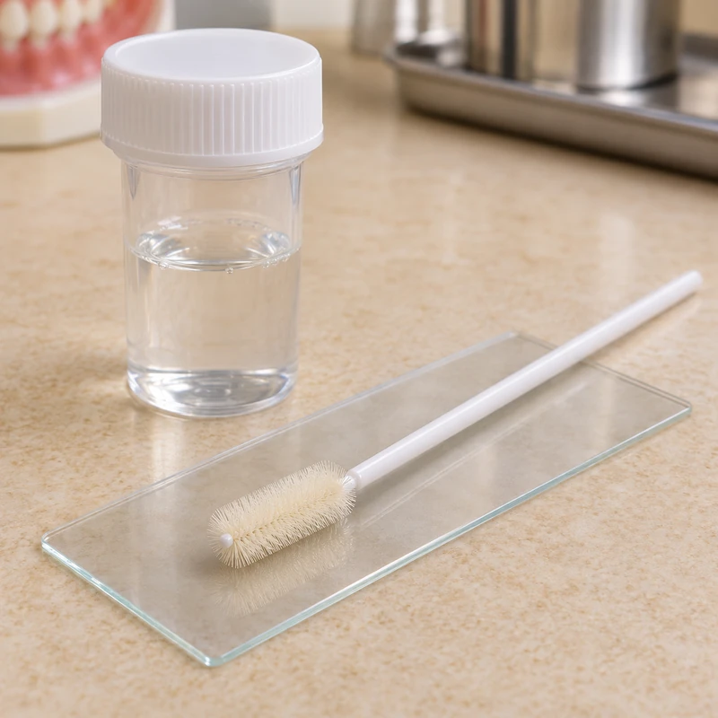
A small brush collects cells from the abnormal area. The choice of investigation depends on the clinical findings.
The laboratory examines the cells for abnormalities. A negative result cannot reliably rule out malignancy.
Suspicious lesions warrant prompt biopsy or specialist referral. A tissue specimen also allows assessment of the tissue layers.
A small brush collects cells from the abnormal area. The choice of investigation depends on the clinical findings.
The laboratory examines the cells for abnormalities. A negative result cannot reliably rule out malignancy.
Suspicious lesions warrant prompt biopsy or specialist referral. A tissue specimen also allows assessment of the tissue layers.
The ADA does not recommend cytological screening of normal oral mucosa.
In specific circumstances when an indicated biopsy cannot currently be performed.
A suspicious or persistent change still requires assessment.
Three steps. No audio.
Each scene can also be selected manually.

Silent film with English on-screen text and simplified teaching images.
A biopsy obtains intact tissue for histological examination.
The clinician inspects the mucosa and feels abnormal areas and adjacent structures as appropriate.
Further assessment is needed. Brush sampling alone cannot establish a final cancer diagnosis.
Report the change and agree the next diagnostic step, even if a previous brush sample was negative.
Ask which investigation the clinical finding needs and when the result will be reviewed.
If the area persists, when will a biopsy be arranged?
A persistent abnormal area needs dental or specialist assessment.
Call the practice · 0221 669 401 80Telephone contactLimited sampling and lack of intact architecture constrain cytology.
Editorial research: . General information, not an individual diagnosis or treatment recommendation. Teaching illustrations are simplified and do not depict a patient case.
If you turn this on, this website stores which guides you open, separately from advertising preferences. The markers expire after a fixed 30-day period from activation. The list is not sent to us by this feature. You can turn it off here and delete the markers.
Other people using the same browser profile can also see the markers. German and English versions share one marker. Expired entries are removed the next time you open a page with this feature; deleting browser data may remove them earlier. “Viewed” means opened, not read or completed.