What does the first recording provide?
A baseline shape for later comparison.

The dental team records tooth surfaces digitally at different times. After the models have been aligned, changes in shape can be compared. Whether a difference represents real tissue loss also depends on scan quality and measurement error.
For training & practice ↓
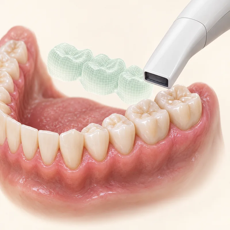
An initial scan records visible surface shape. Flattened edges or hollows can be documented without inferring their cause from shape alone.
The scans must be aligned for comparison. Stable reference surfaces help, while newly placed restorations can affect alignment.
A colour scale can show where surfaces differ. The interval, measurement uncertainty and clinical findings are needed to interpret that difference.
An initial scan records visible surface shape. Flattened edges or hollows can be documented without inferring their cause from shape alone.
The scans must be aligned for comparison. Stable reference surfaces help, while newly placed restorations can affect alignment.
A colour scale can show where surfaces differ. The interval, measurement uncertainty and clinical findings are needed to interpret that difference.
A baseline shape for later comparison.
Reference surface, scale and precision of the method.
It needs to be assessed in relation to the method’s measurement error.
Three steps. No audio.
Each scene can also be selected manually.

Silent film with English on-screen text and simplified teaching images.
For a new crown the current shape matters. Wear monitoring compares shape specifically over time.
It records the current shape. A second comparable recording is needed to measure change.
No. Scanning conditions and alignment can create apparent differences. The scale used by the particular software matters.
It shows differences in shape. Diet, medical history, tooth contacts and other findings help determine the cause.
A question to bring along
Which change in this comparison is larger than the expected measurement uncertainty?
Earlier scans, photographs or models can be helpful at a wear review.
Choose an appointmentAppointment selection at DoctolibIn one evaluated protocol, height measurements were more robust than volume measurements. Results depend on the method and time interval studied.
Editorial research: . General information, not an individual diagnosis or treatment recommendation. Teaching illustrations are simplified and do not depict a patient case.
If you turn this on, this website stores which guides you open, separately from advertising preferences. The markers expire after a fixed 30-day period from activation. The list is not sent to us by this feature. You can turn it off here and delete the markers.
Other people using the same browser profile can also see the markers. German and English versions share one marker. Expired entries are removed the next time you open a page with this feature; deleting browser data may remove them earlier. “Viewed” means opened, not read or completed.