Is looking at the gum enough?
Additional tests are needed to identify the source.

A small opening in the gum can communicate with an inflammatory focus through a tract. A clinician can trace it with a radiopaque gutta-percha point and a targeted radiograph. The tooth nearest the opening is not always the source.
For training & practice ↓
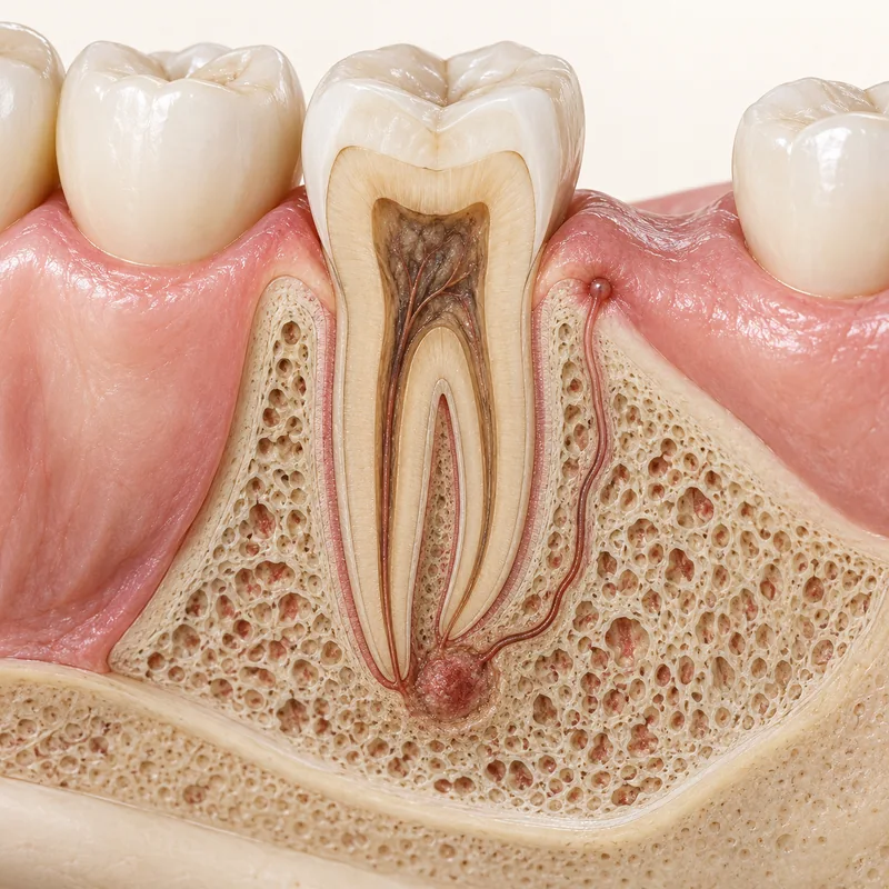
Fluid may drain through a small opening. As the tract can travel sideways, nearby teeth are also examined.
A flexible radiopaque gutta-percha point can mark the path. This is a clinical examination. Do not probe the opening yourself.
This illustration shows two root tips without a marker. To identify the source, the clinician compares the tracing point on the radiograph with findings at the teeth and gums.
Fluid may drain through a small opening. As the tract can travel sideways, nearby teeth are also examined.
A flexible radiopaque gutta-percha point can mark the path. This is a clinical examination. Do not probe the opening yourself.
This illustration shows two root tips without a marker. To identify the source, the clinician compares the tracing point on the radiograph with findings at the teeth and gums.
Additional tests are needed to identify the source.
Relief alone does not confirm healing.
Have the area examined and avoid manipulating it.
Three steps. No audio.
Each scene can also be selected manually.

Silent film with English on-screen text and simplified teaching images.
A marker is placed temporarily in the sinus tract. A root filling is placed inside a cleaned root canal.
No. A prospective study of 100 tracts frequently found that the opening was displaced from the responsible tooth.
Drainage may temporarily relieve pressure. The source may persist even if the opening disappears for a while.
It is used temporarily for tracing. Treatment is planned after the source has been identified.
A question to bring along
Which findings indicate that this particular tooth is the source?
Recurrent openings, pus drainage or swelling warrant prompt assessment.
Choose an appointmentAppointment selection at DoctolibThe two-dimensional image is interpreted alongside the clinical findings.
Editorial research: . General information, not an individual diagnosis or treatment recommendation. Teaching illustrations are simplified and do not depict a patient case.
If you turn this on, this website stores which guides you open, separately from advertising preferences. The markers expire after a fixed 30-day period from activation. The list is not sent to us by this feature. You can turn it off here and delete the markers.
Other people using the same browser profile can also see the markers. German and English versions share one marker. Expired entries are removed the next time you open a page with this feature; deleting browser data may remove them earlier. “Viewed” means opened, not read or completed.