What matters besides brightness?
The relationship to the tooth and its condition.

In condensing osteitis, bone near a tooth root becomes denser in response to a persistent inflammatory stimulus. It may appear lighter on a radiograph. Assessment includes the associated tooth, its history and clinical findings.
For training & practice ↓
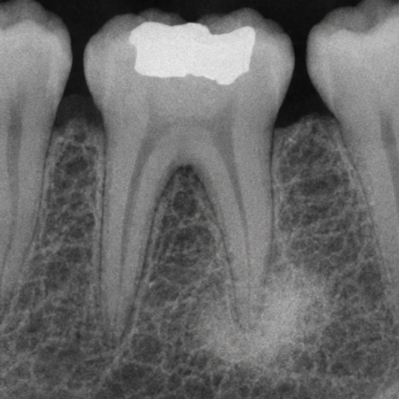
In the simulated radiographic image, bone around one root appears denser than its surroundings. The pattern illustrates a finding rather than establishing a diagnosis.
The cutaway shows the connection between pulp chamber and root canal. Symptoms, sensibility and percussion tests and previous treatment add context.
The denser area may recede or persist at follow-up. Radiographic changes are assessed alongside the condition of the tooth.
In the simulated radiographic image, bone around one root appears denser than its surroundings. The pattern illustrates a finding rather than establishing a diagnosis.
The cutaway shows the connection between pulp chamber and root canal. Symptoms, sensibility and percussion tests and previous treatment add context.
The denser area may recede or persist at follow-up. Radiographic changes are assessed alongside the condition of the tooth.
The relationship to the tooth and its condition.
The combined clinical and radiographic diagnosis.
They make changes easier to recognise.
Three steps. No audio.
Each scene can also be selected manually.

Silent film with English on-screen text and simplified teaching images.
This may also appear as a dense area. An inflammatory relationship with a tooth is central to condensing osteitis.
No. Different conditions appear dense. Association with the tooth and other findings help identify an inflammatory cause.
The indication depends on the tooth’s diagnosis. A dense area alone does not determine treatment.
In a follow-up study many of the investigated roots showed regression of the dense area after endodontic treatment. The pace and outcome vary.
A question to bring along
Which findings suggest this dense area is related to this tooth?
Previous radiographs and a history of earlier treatment can help assessment.
Choose an appointmentAppointment selection at DoctolibSimilar image density can occur in conditions with different causes.
Editorial research: . General information, not an individual diagnosis or treatment recommendation. Teaching illustrations are simplified and do not depict a patient case.
If you turn this on, this website stores which guides you open, separately from advertising preferences. The markers expire after a fixed 30-day period from activation. The list is not sent to us by this feature. You can turn it off here and delete the markers.
Other people using the same browser profile can also see the markers. German and English versions share one marker. Expired entries are removed the next time you open a page with this feature; deleting browser data may remove them earlier. “Viewed” means opened, not read or completed.