Part of the tooth?
No; surrounding bone.

The lamina dura is the dense bony boundary of the tooth socket, often seen on a dental radiograph as a fine bright line around the root. The periodontal ligament space lies between the root surface and this bone wall. Visibility also depends on projection. An indistinct or apparently interrupted line alone therefore does not establish a diagnosis.
For training & practice ↓
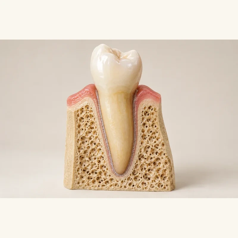
The lamina dura belongs to the tooth-socket wall. It lies outside the darker periodontal ligament space, not on the root surface itself.
Radiographs superimpose three-dimensional structures. The same bone wall may appear sharp, indistinct or barely visible depending on beam direction.
The outline, surrounding bone, symptoms and examination must be considered together. One image feature does not determine treatment.
The lamina dura belongs to the tooth-socket wall. It lies outside the darker periodontal ligament space, not on the root surface itself.
Radiographs superimpose three-dimensional structures. The same bone wall may appear sharp, indistinct or barely visible depending on beam direction.
The outline, surrounding bone, symptoms and examination must be considered together. One image feature does not determine treatment.
No; surrounding bone.
A fine bright line.
No.
Three steps. No audio.
Each scene can also be selected manually.

Silent educational film with German on-screen text and simplified teaching images. Not a patient case or a promised treatment outcome. The full English explanation is available as text.
Cementum covers the root. The lamina dura belongs to the socket wall, with periodontal supporting tissue in the space between them.
No. It belongs to the surrounding jawbone, not to enamel, dentine or root cementum.
The dense socket wall attenuates x-rays. With a suitable projection it appears as a narrow radiopaque line.
No. That space lies between the root and socket wall and usually appears darker. The lamina dura forms its bony boundary.
Not alone. Disease can alter the line, but beam direction and superimposition also affect its appearance. Clinical interpretation is needed.
No. One radiographic feature cannot replace examination or assessment of other imaging findings and symptoms.
You can bring this question to your appointment:
Is this outline truly altered, or could projection explain its appearance?
A dental examination can clarify how this relates to your findings and treatment planning.
Discuss your dental findingsAppointment selection at DoctolibCementum covers the root. The lamina dura belongs to the socket wall, with periodontal supporting tissue in the space between them.
Editorial research: . General information, not an individual diagnosis or treatment recommendation. Teaching illustrations are simplified and do not depict a patient case.
If you turn this on, this website stores which guides you open, separately from advertising preferences. The markers expire after a fixed 30-day period from activation. The list is not sent to us by this feature. You can turn it off here and delete the markers.
Other people using the same browser profile can also see the markers. German and English versions share one marker. Expired entries are removed the next time you open a page with this feature; deleting browser data may remove them earlier. “Viewed” means opened, not read or completed.