Who plans treatment?
Orthodontic and maxillofacial surgery teams coordinate care.

In a Le Fort I osteotomy, the surgical team separates the tooth-bearing upper jaw along a planned level. The bone moves with the upper teeth and is fixed in the planned position.
For training & practice ↓
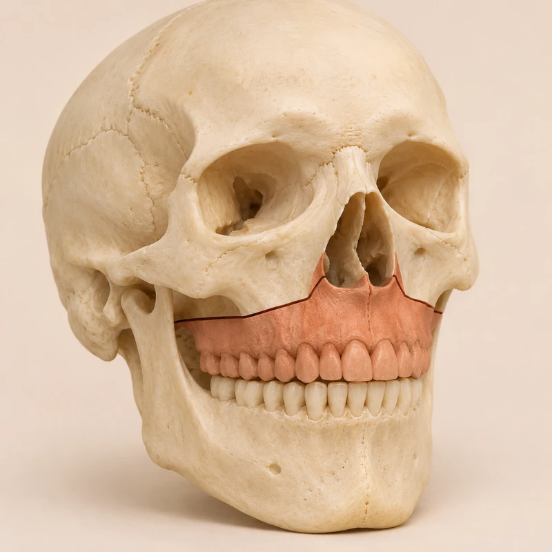
The roots of the upper teeth sit in the tooth-bearing part. During repositioning this section moves as a bony unit.
Planning takes account of how the arches meet and how the upper jaw sits in the face. The direction of movement is individual.
Small plates and screws can secure the new bone position during healing. Follow-up assesses the bite, wounds and recovery.
The roots of the upper teeth sit in the tooth-bearing part. During repositioning this section moves as a bony unit.
Planning takes account of how the arches meet and how the upper jaw sits in the face. The direction of movement is individual.
Small plates and screws can secure the new bone position during healing. Follow-up assesses the bite, wounds and recovery.
Orthodontic and maxillofacial surgery teams coordinate care.
The approach is usually inside the mouth above the upper teeth.
It depends on the jaw discrepancy.
Three steps. No audio.
Each scene can also be selected manually.

Silent film with English on-screen text and simplified teaching images.
An osteotomy is a planned division of bone. A Le Fort fracture describes an injury pattern after trauma.
Braces mainly move teeth within the jaw. A pronounced skeletal discrepancy may also call for moving the jaw base.
Some plans involve the upper jaw alone. Others call for a combined procedure to achieve the intended bite.
Discuss the planned movement, possible changes in sensation, bleeding, diet during recovery and follow-up for your operation.
A question to bring along
How much of my bite problem comes from tooth position and how much from the jaw base?
Discuss the surgical plan with your orthodontic and maxillofacial teams.
Choose an appointmentAppointment selection at DoctolibIndividual tooth positions and the position of the jaw base are two distinct aspects of planning.
Editorial research: . General information, not an individual diagnosis or treatment recommendation. Teaching illustrations are simplified and do not depict a patient case.
If you turn this on, this website stores which guides you open, separately from advertising preferences. The markers expire after a fixed 30-day period from activation. The list is not sent to us by this feature. You can turn it off here and delete the markers.
Other people using the same browser profile can also see the markers. German and English versions share one marker. Expired entries are removed the next time you open a page with this feature; deleting browser data may remove them earlier. “Viewed” means opened, not read or completed.