Can a photo confirm dysplasia?
The diagnosis depends on microscopic examination of tissue.

Oral epithelial dysplasia is an abnormality in cells and their arrangement in the surface lining of the mouth, seen under a microscope. It can increase the risk of oral cancer. It is distinct from invasive cancer.
For training & practice ↓
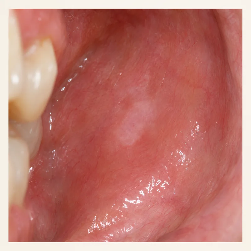
Some areas appear white or red. Colour and texture may prompt an examination but cannot reliably reveal cellular changes.
When indicated, a clinician takes a small tissue sample. The pathologist examines cell shape, maturation and arrangement in the epithelium.
Ask what grade the pathologist found, whether further treatment is recommended and when your next review is due.
Some areas appear white or red. Colour and texture may prompt an examination but cannot reliably reveal cellular changes.
When indicated, a clinician takes a small tissue sample. The pathologist examines cell shape, maturation and arrangement in the epithelium.
Ask what grade the pathologist found, whether further treatment is recommended and when your next review is due.
The diagnosis depends on microscopic examination of tissue.
Outcomes vary. Individual follow-up is important.
The pathology report refers to the area actually sampled.
Three steps. No audio.
Each scene can also be selected manually.

Silent film with English on-screen text and simplified teaching images.
Leukoplakia describes a clinically defined white patch. Dysplasia describes tissue changes.
The laboratory examines the submitted tissue. The clinician also considers the extent, location and rest of the mouth.
Keep the scheduled review. Symptoms tell little about the cellular findings.
Contact the team sooner if a known patch grows, bleeds or becomes firm.
A question to bring along
How does the grade affect my care?
Have an unexplained oral lesion assessed and arrange the recommended review.
Choose an appointmentAppointment selection at DoctolibClinical description and pathology answer different questions about the same area.
Editorial research: . General information, not an individual diagnosis or treatment recommendation. Teaching illustrations are simplified and do not depict a patient case.
If you turn this on, this website stores which guides you open, separately from advertising preferences. The markers expire after a fixed 30-day period from activation. The list is not sent to us by this feature. You can turn it off here and delete the markers.
Other people using the same browser profile can also see the markers. German and English versions share one marker. Expired entries are removed the next time you open a page with this feature; deleting browser data may remove them earlier. “Viewed” means opened, not read or completed.