Where is the finding?
In tissues around the root tip.

A periapical granuloma is inflamed tissue rich in cells and small blood vessels around a root tip, often in response to infection in the canal system. A dark area on an X-ray alone cannot identify the tissue with certainty.
For training & practice ↓
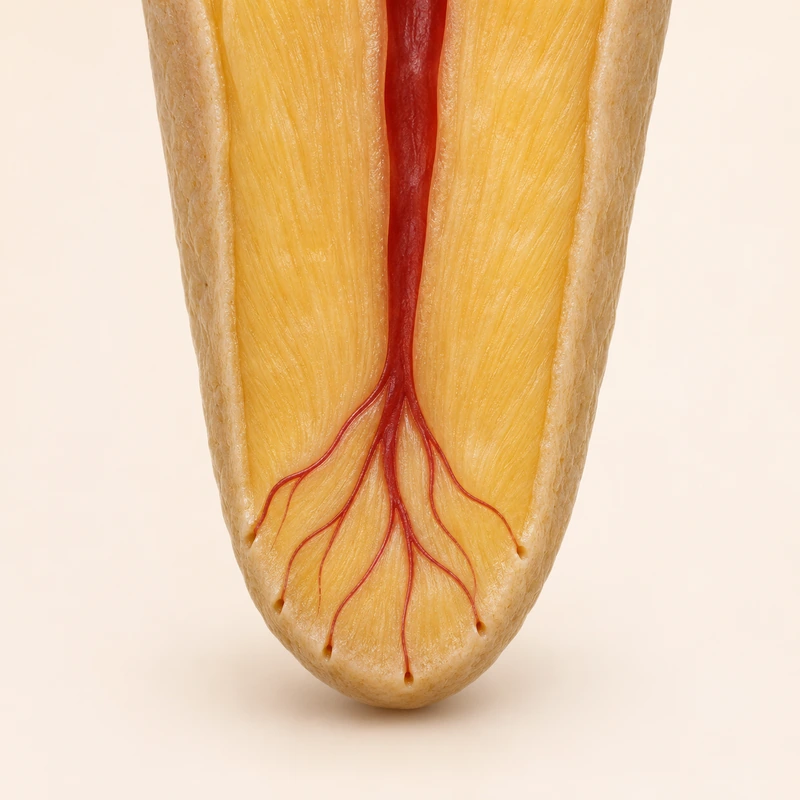
The root canal communicates with surrounding tissues near its tip. Infection in the canal can trigger inflammation in this region.
A dark area can indicate local bone loss. Shape and size alone cannot reliably separate a granuloma from a cyst.
When tissue is removed for clinical reasons, examination under the microscope can determine the nature of the lesion.
The root canal communicates with surrounding tissues near its tip. Infection in the canal can trigger inflammation in this region.
A dark area can indicate local bone loss. Shape and size alone cannot reliably separate a granuloma from a cyst.
When tissue is removed for clinical reasons, examination under the microscope can determine the nature of the lesion.
In tissues around the root tip.
Histological assessment of an appropriate tissue specimen.
Whether the associated tooth is the source.
Three steps. No audio.
Each scene can also be selected manually.

Silent film with English on-screen text and simplified teaching images.
A radicular cyst has a cavity lined by epithelium. A periapical granuloma consists of inflamed tissue.
Changes at a root tip may go unnoticed for some time. The assessment includes pulp tests and the tooth’s treatment history.
History, examination and follow-up often guide treatment of a suspected endodontic cause. Atypical or persistent findings may need further investigation.
If the canal is infected, treatment targets that source. Initial treatment, retreatment or another approach may be appropriate depending on the tooth and the findings.
A question to bring along
Which tooth might be the source, and how will healing be followed?
Have an unexplained finding compared with your earlier images.
Choose an appointmentAppointment selection at DoctolibA swelling on the gum and a granuloma around a root tip describe different findings. The site of origin helps distinguish them.
Editorial research: . General information, not an individual diagnosis or treatment recommendation. Teaching illustrations are simplified and do not depict a patient case.
If you turn this on, this website stores which guides you open, separately from advertising preferences. The markers expire after a fixed 30-day period from activation. The list is not sent to us by this feature. You can turn it off here and delete the markers.
Other people using the same browser profile can also see the markers. German and English versions share one marker. Expired entries are removed the next time you open a page with this feature; deleting browser data may remove them earlier. “Viewed” means opened, not read or completed.