Which tissue is treated?
The surface layer of the enamel.

In enamel microabrasion, a dentist uses an acid-abrasive paste to remove a thin surface layer of enamel. Superficial patches can become less noticeable. The depth of a mark limits how much its appearance can change.
For training & practice ↓
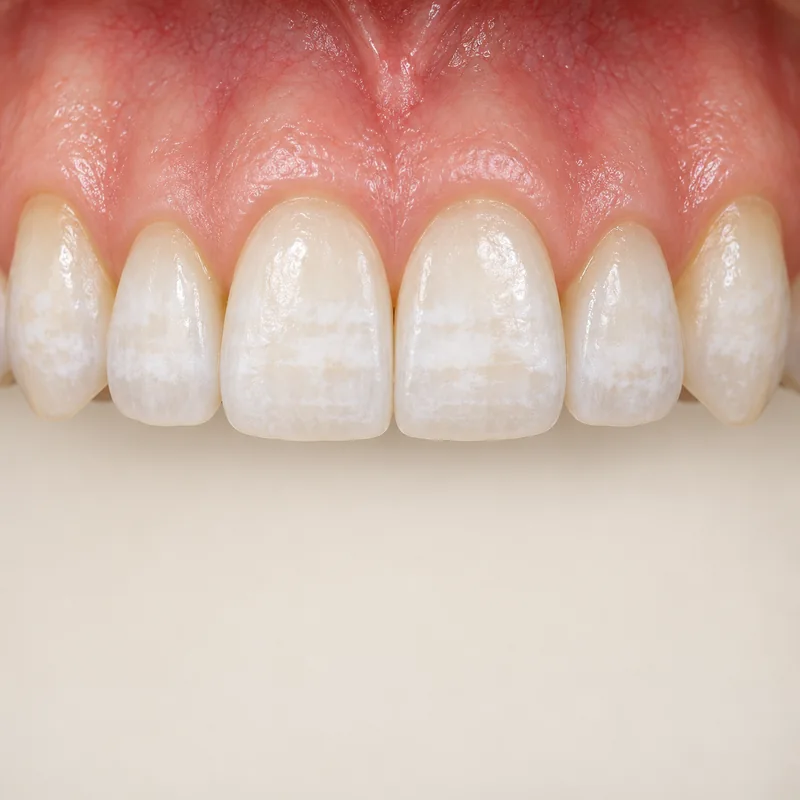
Pale patches may extend to different depths. An examination first assesses their cause and position.
An acid-abrasive paste acts on the outer enamel. The tissue removed is not replaced by polishing.
After treatment, the dentist compares colour and surface texture. Deeper changes can remain visible.
Pale patches may extend to different depths. An examination first assesses their cause and position.
An acid-abrasive paste acts on the outer enamel. The tissue removed is not replaced by polishing.
After treatment, the dentist compares colour and surface texture. Deeper changes can remain visible.
The surface layer of the enamel.
Discolouration extending deeper into enamel or dentine.
Enamel that has been removed does not grow back.
Three steps. No audio.
Each scene can also be selected manually.

Silent film with English on-screen text and simplified teaching images.
Microabrasion removes surface tissue. Bleaching uses peroxides to change pigments chemically. A mark that responds to one approach may respond differently to the other.
White patches may reflect fluorosis, developmental defects or early decay. If decay is active, controlling the disease takes priority.
Cleaning removes deposits from a tooth. Microabrasion targets a patch within the superficial tooth tissue itself.
The dentist assesses colour, extent, texture and depth. Comparing the area under similar lighting helps assess the change.
When discussing white or brown enamel marks
What suggests that my enamel mark is superficial?
Have the cause of an enamel mark assessed before cosmetic treatment.
Choose an appointmentAppointment selection at DoctolibResin infiltration changes how light travels through porous enamel by introducing resin. Microabrasion acts mainly by shallow tissue removal.
Editorial research: . General information, not an individual diagnosis or treatment recommendation. Teaching illustrations are simplified and do not depict a patient case.
If you turn this on, this website stores which guides you open, separately from advertising preferences. The markers expire after a fixed 30-day period from activation. The list is not sent to us by this feature. You can turn it off here and delete the markers.
Other people using the same browser profile can also see the markers. German and English versions share one marker. Expired entries are removed the next time you open a page with this feature; deleting browser data may remove them earlier. “Viewed” means opened, not read or completed.