Which root?
The front root on the cheek side of an upper molar.

MB2 is a second canal in the front, cheek-side root of an upper molar. Two canals can remain separate within the same root or join further along.
For training & practice ↓
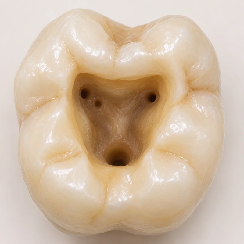
Through an opening in the biting surface, the entrances in the pulp chamber floor come into view. Two nearby openings may lead into the same root.
The extra canal entrance can be difficult to locate by eye. Magnification and lighting help the clinician interpret the shape of the chamber floor.
An infected canal left untreated can sustain inflammation around the root tip. Other causes also need to be considered before retreatment.
Through an opening in the biting surface, the entrances in the pulp chamber floor come into view. Two nearby openings may lead into the same root.
The extra canal entrance can be difficult to locate by eye. Magnification and lighting help the clinician interpret the shape of the chamber floor.
An infected canal left untreated can sustain inflammation around the root tip. Other causes also need to be considered before retreatment.
The front root on the cheek side of an upper molar.
By inspecting the chamber floor and using additional imaging where appropriate.
If symptoms or changes around the root tip persist.
Three steps. No audio.
Each scene can also be selected manually.

Silent film with English on-screen text and simplified teaching images.
Two entrances at the chamber floor may join along the root to form one path.
It describes an additional canal within the mesiobuccal root. Root count and canal count describe different things.
Examination and conventional images are the starting point. Whether a small-volume 3D scan could answer an unresolved question is assessed individually.
Anatomy varies. A missed canal is one possible explanation for persistent symptoms or a lesion that fails to heal. Symptoms alone do not prove it.
A question to bring along
What findings suggest an additional canal in this tooth?
Bring images and details of any earlier root canal treatment for review.
Choose an appointmentAppointment selection at DoctolibA retrospective imaging study found an association between untreated MB2 canals and apical changes. Imaging alone cannot establish the cause in an individual tooth.
Editorial research: . General information, not an individual diagnosis or treatment recommendation. Teaching illustrations are simplified and do not depict a patient case.
If you turn this on, this website stores which guides you open, separately from advertising preferences. The markers expire after a fixed 30-day period from activation. The list is not sent to us by this feature. You can turn it off here and delete the markers.
Other people using the same browser profile can also see the markers. German and English versions share one marker. Expired entries are removed the next time you open a page with this feature; deleting browser data may remove them earlier. “Viewed” means opened, not read or completed.