Can it be benign and still cause harm?
Yes. Local growth can damage bone and adjacent structures.

An ameloblastoma is an uncommon tumour arising from tooth-forming epithelium. It is usually benign but can grow into adjacent jawbone and displace or destroy it. The back of the lower jaw is a common site.
For training & practice ↓
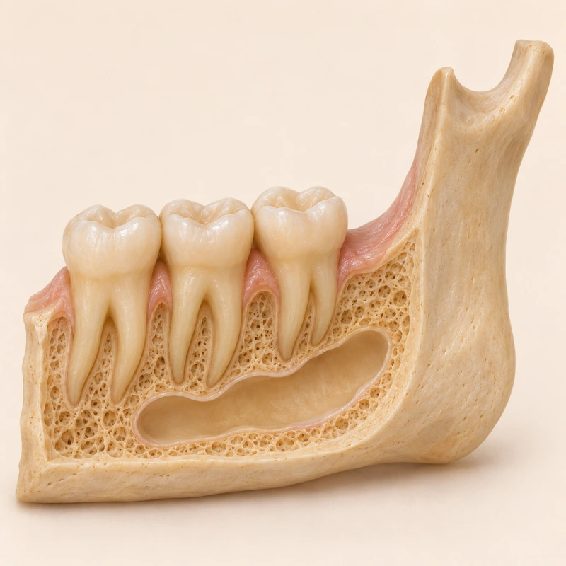
A slow-growing, initially painless swelling may be the first change noticed. Sometimes a lesion appears on imaging obtained for another reason.
Imaging shows location and extent. A dark area with one or several compartments can have various causes, including a cyst.
Examination of tissue helps establish the diagnosis. Long-term review matters because an ameloblastoma can recur in the same area.
A slow-growing, initially painless swelling may be the first change noticed. Sometimes a lesion appears on imaging obtained for another reason.
Imaging shows location and extent. A dark area with one or several compartments can have various causes, including a cyst.
Examination of tissue helps establish the diagnosis. Long-term review matters because an ameloblastoma can recur in the same area.
Yes. Local growth can damage bone and adjacent structures.
An X-ray alone does not identify the tissue type.
Oral and maxillofacial specialists assess the lesion and plan care.
Three steps. No audio.
Each scene can also be selected manually.

Silent film with English on-screen text and simplified teaching images.
A cyst forms a cavity. Some tumours can also look like cavities on imaging.
It can show the extent of the lesion within bone and its relationship to nearby structures.
The approach depends on tissue diagnosis, location and extent. A specialist team discusses the options.
Recurrence can happen later. Keep the follow-up schedule agreed with your team.
A question to bring along
Who coordinates tissue diagnosis and follow-up?
Seek specialist assessment for persistent jaw swelling or an unexplained bone lesion.
Choose an appointmentAppointment selection at DoctolibBenign describes biological classification. Local behaviour also influences the extent of treatment.
Editorial research: . General information, not an individual diagnosis or treatment recommendation. Teaching illustrations are simplified and do not depict a patient case.
If you turn this on, this website stores which guides you open, separately from advertising preferences. The markers expire after a fixed 30-day period from activation. The list is not sent to us by this feature. You can turn it off here and delete the markers.
Other people using the same browser profile can also see the markers. German and English versions share one marker. Expired entries are removed the next time you open a page with this feature; deleting browser data may remove them earlier. “Viewed” means opened, not read or completed.