Where is it?
Close to the root tip.

An apical delta is a fine branching network of the root canal system close to the root tip. Several small connections may lead to the surrounding tissues instead of a single simple exit. This anatomical variation does not itself diagnose inflammation, but may make an infected canal system harder to clean.
For training & practice ↓
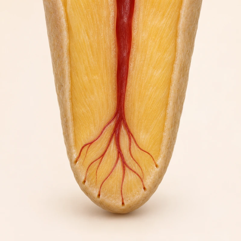
The small branches occupy the end portion of the root. Their shape, number and course vary; a teaching image shows only a simplified example.
Files cannot fully instrument tiny curved connections. Mechanical preparation is therefore combined with chemical cleaning.
An apical delta alone does not justify an intervention. If disease is present, the overall findings and response to treatment guide management.
The small branches occupy the end portion of the root. Their shape, number and course vary; a teaching image shows only a simplified example.
Files cannot fully instrument tiny curved connections. Mechanical preparation is therefore combined with chemical cleaning.
An apical delta alone does not justify an intervention. If disease is present, the overall findings and response to treatment guide management.
Close to the root tip.
No. Fine branches can be beyond imaging resolution.
Adapted treatment and assessment of healing over time.
Three steps. No audio.
Each scene can also be selected manually.

Silent educational film with German on-screen text and simplified teaching images. Not a patient case or a promised treatment outcome. The full English explanation is available as text.
A lateral canal is an additional connection from the side of a main canal. In an apical delta, the terminal canal region divides into several small branches. These terms describe different arrangements.
No. Branches may be smaller than structures that can be resolved reliably. Even CBCT does not always show these details fully.
No. Anatomy alone is not a surgical indication. Apical surgery is considered only after an appropriate individual assessment.
No. Radiopaque material is not direct proof that all tissue remnants or microbes were removed. Clinical progress and follow-up still matter.
You can bring this question to your appointment:
How does the branching near the root tip affect treatment planning and follow-up?
An individual examination can clarify the findings and suitable next steps.
Arrange an assessmentAppointment selection at DoctolibA lateral canal is an additional connection from the side of a main canal. In an apical delta, the terminal canal region divides into several small branches. These terms describe different arrangements.
Editorial research: . General information, not an individual diagnosis or treatment recommendation. Teaching illustrations are simplified and do not depict a patient case.
If you turn this on, this website stores which guides you open, separately from advertising preferences. The markers expire after a fixed 30-day period from activation. The list is not sent to us by this feature. You can turn it off here and delete the markers.
Other people using the same browser profile can also see the markers. German and English versions share one marker. Expired entries are removed the next time you open a page with this feature; deleting browser data may remove them earlier. “Viewed” means opened, not read or completed.