Does optical capture use X-rays?
The optical surface scan itself uses no X-rays.

A digital facial scan records the outer shape of the face in three dimensions. Optical images produce a surface model. It can be matched to a separate scan of the teeth to assess features such as the dental midline and smile line in the context of the lips and face.
For training & practice ↓
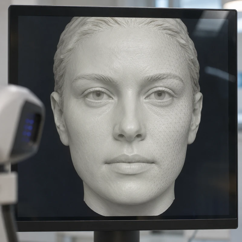
The device captures visible contours. A neutral expression or a smile is recorded to answer the clinical question. Lip movement changes what is seen.
A separate intraoral scan provides tooth-surface detail. Combining the two records requires shared reference features such as visible teeth or suitable markers.
If matching is inaccurate, the teeth can appear shifted in the virtual face. The team checks features such as the midline and the inclination of the biting plane.
The device captures visible contours. A neutral expression or a smile is recorded to answer the clinical question. Lip movement changes what is seen.
A separate intraoral scan provides tooth-surface detail. Combining the two records requires shared reference features such as visible teeth or suitable markers.
If matching is inaccurate, the teeth can appear shifted in the virtual face. The team checks features such as the midline and the inclination of the biting plane.
The optical surface scan itself uses no X-rays.
Aligning separate records using features they have in common.
The simulation supports planning and discussion of your preferences.
Three steps. No audio.
Each scene can also be selected manually.

Silent film with English on-screen text and simplified teaching images.
This uses dedicated markers to record implant positions. A facial scan focuses on external soft tissue.
It records the surface using light. It cannot answer questions about the inside of bone or root tips. Those findings may call for different investigations.
Cheeks and lips obscure much of the dentition. An intraoral scan adds the visible shape of the teeth and allows matching with the facial record.
A comparison found differences according to scanner and matching method. An attractive-looking model may still contain small positional errors.
A question to bring along
What question will the scan answer, and how is its alignment with my dental model checked?
Ask which record represents the lips and which represents the teeth when discussing a digital plan.
Choose a dental appointmentAppointment selection at DoctolibSurface detail and accuracy of registration are separate measurement tasks.
Editorial research: . General information, not an individual diagnosis or treatment recommendation. Teaching illustrations are simplified and do not depict a patient case.
If you turn this on, this website stores which guides you open, separately from advertising preferences. The markers expire after a fixed 30-day period from activation. The list is not sent to us by this feature. You can turn it off here and delete the markers.
Other people using the same browser profile can also see the markers. German and English versions share one marker. Expired entries are removed the next time you open a page with this feature; deleting browser data may remove them earlier. “Viewed” means opened, not read or completed.