Should I attend if it feels fine?
Keep the follow-up appointments arranged after the injury.

A serious injury can damage the protective surface of the root. If the tissue inside the canal also dies and becomes infected, an inflammatory response can sustain loss of root tissue from the outside.
For training & practice ↓
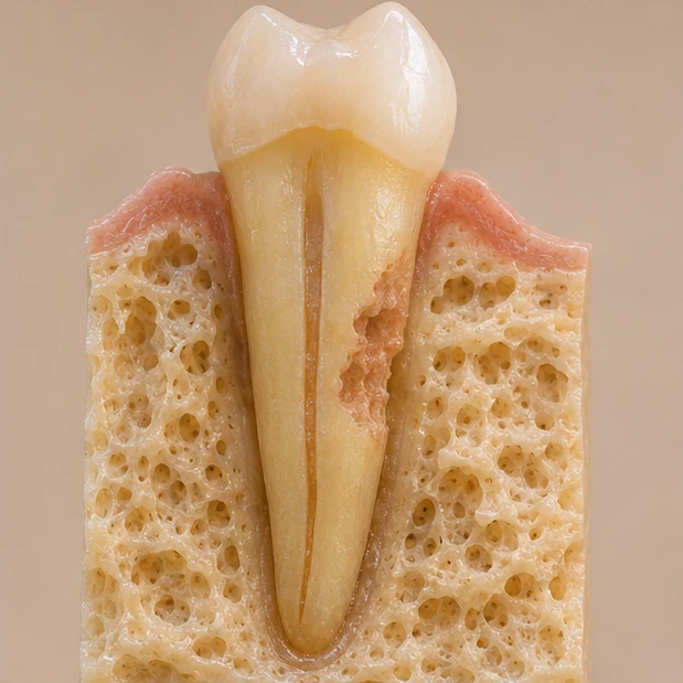
Displacement can injure both the tooth’s supporting tissues and its outer root covering, leaving a surface vulnerable to resorbing cells.
Infected tissue inside can maintain inflammation at the damaged outer surface. Assessment therefore considers the pulp as well as the root defect.
Comparable radiographs may reveal a new or enlarging defect. Confirmed infection-related resorption warrants a prompt endodontic decision.
Displacement can injure both the tooth’s supporting tissues and its outer root covering, leaving a surface vulnerable to resorbing cells.
Infected tissue inside can maintain inflammation at the damaged outer surface. Assessment therefore considers the pulp as well as the root defect.
Comparable radiographs may reveal a new or enlarging defect. Confirmed infection-related resorption warrants a prompt endodontic decision.
Keep the follow-up appointments arranged after the injury.
Several findings are assessed together after trauma.
Treat the source of infection and limit further resorption.
Three steps. No audio.
Each scene can also be selected manually.

Silent film with English on-screen text and simplified teaching images.
Bone replaces root structure after severe injury to the supporting tissues. These processes can require different management after trauma.
Yes. An injured tooth may feel comfortable while its root changes. The scheduled trauma follow-ups still matter.
The response may temporarily disappear after trauma. Other findings and follow-up help identify pulp death.
When the canal is infected, treatment aims to remove the inflammatory stimulus and arrest further loss. It does not regenerate missing root structure.
A question to bring along
How has my root changed since the last radiograph?
Follow-up visits are part of care after tooth displacement.
Choose an appointmentAppointment selection at DoctolibThe injury history, location and change over time help interpret a small dark area.
Editorial research: . General information, not an individual diagnosis or treatment recommendation. Teaching illustrations are simplified and do not depict a patient case.
If you turn this on, this website stores which guides you open, separately from advertising preferences. The markers expire after a fixed 30-day period from activation. The list is not sent to us by this feature. You can turn it off here and delete the markers.
Other people using the same browser profile can also see the markers. German and English versions share one marker. Expired entries are removed the next time you open a page with this feature; deleting browser data may remove them earlier. “Viewed” means opened, not read or completed.