Must there be a visible spot?
A change in colour can be absent.

External cervical resorption is the loss of hard tooth tissue by the body’s own cells, starting at the outer surface near the neck of the tooth. It can spread beneath the surface and around the root.
For training & practice ↓
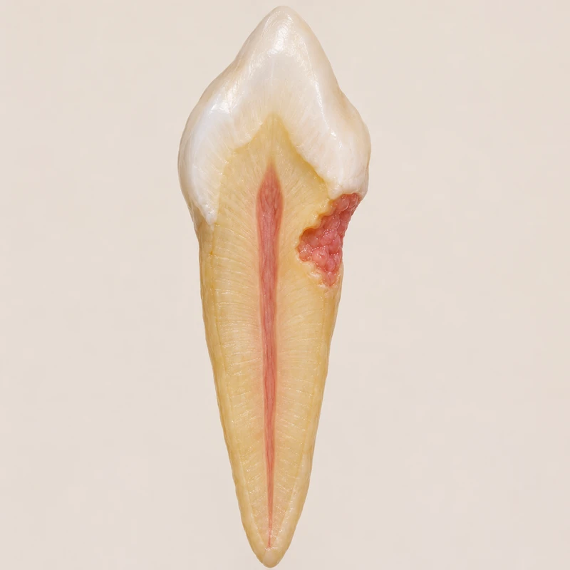
The entry point can be small and close to the gum. Loss beneath the surface may extend beyond the visible opening.
The process starts externally. A thin tissue layer can initially protect the pulp even when the lesion overlaps the canal on a radiograph.
Repair depends on reaching the affected parts and on sufficient remaining root structure. Follow-up is part of the plan.
The entry point can be small and close to the gum. Loss beneath the surface may extend beyond the visible opening.
The process starts externally. A thin tissue layer can initially protect the pulp even when the lesion overlaps the canal on a radiograph.
Repair depends on reaching the affected parts and on sufficient remaining root structure. Follow-up is part of the plan.
A change in colour can be absent.
On the outer surface at the neck of the tooth.
The extent and accessibility of the defect.
Three steps. No audio.
Each scene can also be selected manually.

Silent film with English on-screen text and simplified teaching images.
In decay, acids produced in plaque dissolve minerals. In resorption, cells in the body remove tooth tissue. Both can occur near the gumline.
A pink hue is possible but is not a reliable test. Many lesions are found on examination or imaging without an obvious colour change.
Pulp status, lesion position and the planned repair guide that decision. An early external defect can occur while the pulp is still vital.
If conventional views leave important questions open, a scan can show how far the lesion extends around and into the root. This can affect the decision about retention.
A question to bring along
How close is the lesion to the canal, and can it be reached for repair?
Arrange an assessment of a newly noticed hollow or colour change at the gumline.
Choose an appointmentAppointment selection at DoctolibA wear notch also often lies near the gumline. Its external outline alone does not establish its cause.
Editorial research: . General information, not an individual diagnosis or treatment recommendation. Teaching illustrations are simplified and do not depict a patient case.
If you turn this on, this website stores which guides you open, separately from advertising preferences. The markers expire after a fixed 30-day period from activation. The list is not sent to us by this feature. You can turn it off here and delete the markers.
Other people using the same browser profile can also see the markers. German and English versions share one marker. Expired entries are removed the next time you open a page with this feature; deleting browser data may remove them earlier. “Viewed” means opened, not read or completed.