What does the device use?
The method described uses optical light.

An optical device illuminates a tooth surface and detects light emitted at a different wavelength. In laser fluorescence, bacterial products can contribute to the signal. The reading adds information to the examination of the surface.
For training & practice ↓
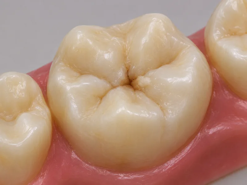
The dental team first examines the clean tooth surface. Natural grooves on a chewing surface may appear dark even without decay.
An optical tip picks up the signal at the examined site. The device and the condition of the surface influence interpretation.
The reading is interpreted alongside the surface appearance, location and caries risk. A radiograph can add information when clinically indicated.
The dental team first examines the clean tooth surface. Natural grooves on a chewing surface may appear dark even without decay.
An optical tip picks up the signal at the examined site. The device and the condition of the surface influence interpretation.
The reading is interpreted alongside the surface appearance, location and caries risk. A radiograph can add information when clinically indicated.
The method described uses optical light.
A single figure is insufficient.
Readings from extracted molars were compared with tissue sections.
Three steps. No audio.
Each scene can also be selected manually.

Silent film with English on-screen text and simplified teaching images.
A curing light hardens restorative material. Diagnostic light is used here to obtain a signal.
No. A fluorescence device uses light. Bitewing radiographs address other questions, such as suspected decay between teeth that cannot easily be inspected.
No. Deposits, staining and measurement conditions can complicate interpretation. Clinical findings, depth and lesion activity guide the decision.
No. In a study of extracted molars the result did not agree with the tissue examination at every site. Laboratory performance cannot be directly transferred to every clinical setting.
A question to bring along
How does the reading fit with the appearance and accessibility of this site?
An uncertain change on a chewing surface can be assessed at an examination.
Choose an appointmentAppointment selection at DoctolibA laboratory comparison with tissue sections can identify errors but cannot predict an individual outcome.
Editorial research: . General information, not an individual diagnosis or treatment recommendation. Teaching illustrations are simplified and do not depict a patient case.
If you turn this on, this website stores which guides you open, separately from advertising preferences. The markers expire after a fixed 30-day period from activation. The list is not sent to us by this feature. You can turn it off here and delete the markers.
Other people using the same browser profile can also see the markers. German and English versions share one marker. Expired entries are removed the next time you open a page with this feature; deleting browser data may remove them earlier. “Viewed” means opened, not read or completed.