What is the anatomical tip of the root called?
The apex.

The apical foramen is the main opening of a root canal into the tissues near the root tip. In a vital tooth it provides a route for vessels and nerve fibres between the pulp and surrounding tissues. It can lie to one side of the anatomical tip.
For training & practice ↓
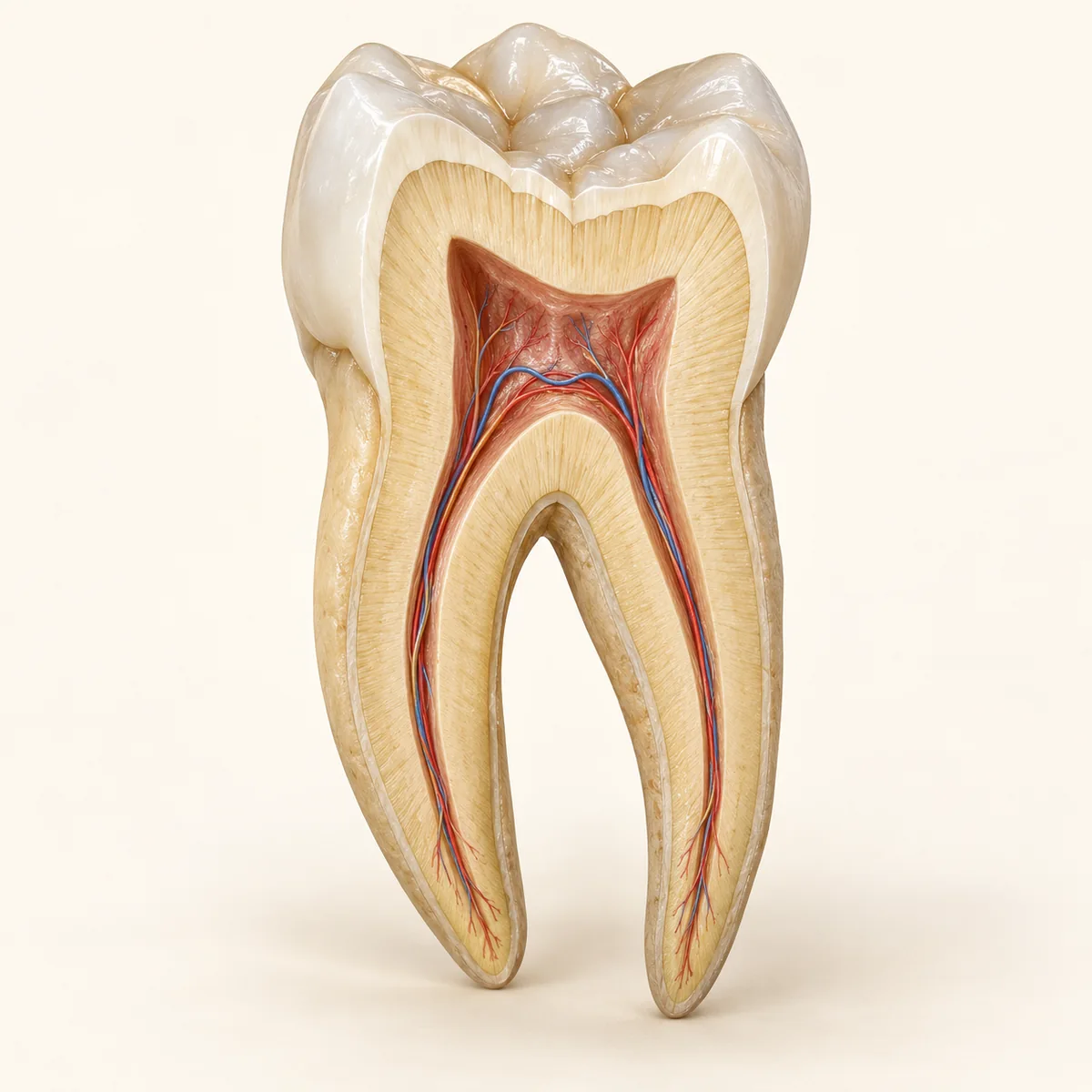
The apex is the anatomical tip of the root. It is a geometric landmark. The actual canal exit need not coincide with this point.
At the opening, the canal communicates with tissues outside the root. Smaller accessory exits may also be present.
The apical constriction is the narrowest part of the canal near its end, when present. The narrowest cross-section and the external opening are distinct landmarks.
The apex is the anatomical tip of the root. It is a geometric landmark. The actual canal exit need not coincide with this point.
At the opening, the canal communicates with tissues outside the root. Smaller accessory exits may also be present.
The apical constriction is the narrowest part of the canal near its end, when present. The narrowest cross-section and the external opening are distinct landmarks.
The apex.
The main opening of the root canal.
The apical constriction.
Three steps. No audio.
Each scene can also be selected manually.

Silent educational film with German on-screen text and simplified teaching images. Not a patient case or a promised treatment outcome. The full English explanation is available as text.
Working length is established for an individual canal from a reference point to the intended end point of treatment. A single point on an image does not determine it.
The apex is the anatomical tip of the root. It is a geometric landmark. The actual canal exit need not coincide with this point.
At the opening, the canal communicates with tissues outside the root. Smaller accessory exits may also be present.
The apical constriction is the narrowest part of the canal near its end, when present. The narrowest cross-section and the external opening are distinct landmarks.
The projected root tip does not necessarily show the exact exit. Additional information is used to establish landmarks during root canal treatment.
Working length is established for an individual canal from a reference point to the intended end point of treatment. A single point on an image does not determine it.
A question you could ask
How did you determine the intended canal end point for my tooth?
When discussing root canal treatment, ask how imaging and electronic length measurement inform the assessment.
Discuss at a dental appointmentAppointment selection at DoctolibWorking length is established for an individual canal from a reference point to the intended end point of treatment. A single point on an image does not determine it.
Editorial research: . General information, not an individual diagnosis or treatment recommendation. Teaching illustrations are simplified and do not depict a patient case.
If you turn this on, this website stores which guides you open, separately from advertising preferences. The markers expire after a fixed 30-day period from activation. The list is not sent to us by this feature. You can turn it off here and delete the markers.
Other people using the same browser profile can also see the markers. German and English versions share one marker. Expired entries are removed the next time you open a page with this feature; deleting browser data may remove them earlier. “Viewed” means opened, not read or completed.