What does the image show?
Differences in X-ray attenuation.

The dentist compares the same tooth surface on images taken at different times. A dark zone extending further into the tooth on comparable projections suggests advancing mineral loss. Clinical examination is also needed to assess whether the surface has broken down.
For training & practice ↓
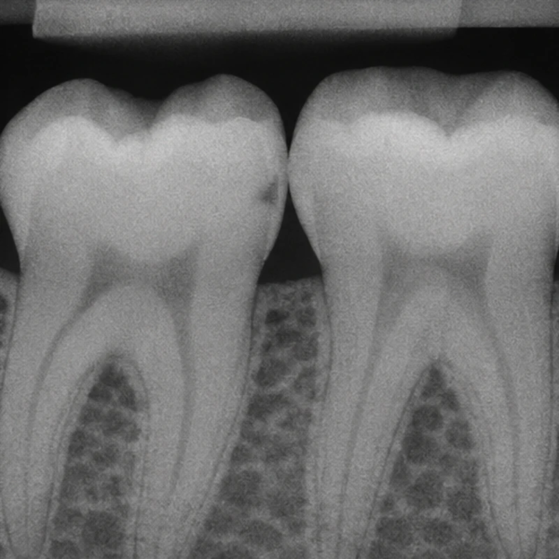
The simulated view shows a small dark patch below the contact between two teeth. Its surface and depth provide a reference for comparison.
The dark area extends further inwards in the second illustration. A change in projection or overlapping contacts can make comparison difficult.
Symptoms, surface integrity and individual risk influence the next step. These illustrations do not document a real patient’s progress.
The simulated view shows a small dark patch below the contact between two teeth. Its surface and depth provide a reference for comparison.
The dark area extends further inwards in the second illustration. A change in projection or overlapping contacts can make comparison difficult.
Symptoms, surface integrity and individual risk influence the next step. These illustrations do not document a real patient’s progress.
Differences in X-ray attenuation.
Individual circumstances and the clinical question.
The same surface at different times.
Three steps. No audio.
Each scene can also be selected manually.

Silent film with English on-screen text and simplified teaching images.
Activity is assessed using clinical features among other findings. Serial images document change between two examinations.
Not necessarily. Radiographs show differences in mineral content. Examination of the accessible surface adds information.
Timing depends on risk, the previous finding and whether the result would influence management.
With a similar projection the dentist can identify changes from baseline. A single exposure cannot show this time course.
A question to bring along
Has this particular lesion changed since my last image?
If available, bring earlier images or the contact details of the practice that took them.
Choose an appointmentAppointment selection at DoctolibRadiographic depth and surface integrity contribute different information to treatment decisions.
Editorial research: . General information, not an individual diagnosis or treatment recommendation. Teaching illustrations are simplified and do not depict a patient case.
If you turn this on, this website stores which guides you open, separately from advertising preferences. The markers expire after a fixed 30-day period from activation. The list is not sent to us by this feature. You can turn it off here and delete the markers.
Other people using the same browser profile can also see the markers. German and English versions share one marker. Expired entries are removed the next time you open a page with this feature; deleting browser data may remove them earlier. “Viewed” means opened, not read or completed.