Is this molar-incisor hypomineralisation?
No. MIH affects enamel formation.

A molar-incisor pattern describes periodontitis in which first permanent molars and incisors preferentially lose support. It can occur in young people. Stage, distribution and evidence of the rate of progression are assessed separately.
For training & practice ↓
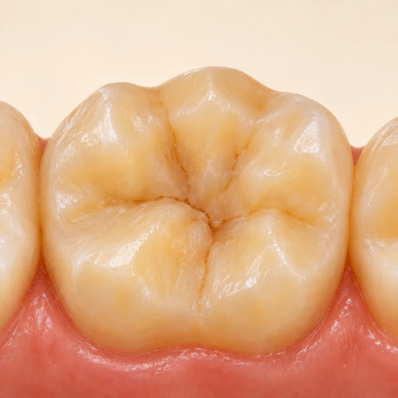
First permanent molars erupt behind the primary teeth. Their broad chewing surfaces are easy to recognise. The periodontal finding affects support below the gum margin.
The clinician also examines the front teeth and compares the findings with the rest of the dentition. Tooth shape alone is not diagnostic.
Probing, clinical attachment loss and appropriate radiographs show which teeth are affected. Previous records may help establish change over time.
First permanent molars erupt behind the primary teeth. Their broad chewing surfaces are easy to recognise. The periodontal finding affects support below the gum margin.
The clinician also examines the front teeth and compares the findings with the rest of the dentition. Tooth shape alone is not diagnostic.
Probing, clinical attachment loss and appropriate radiographs show which teeth are affected. Previous records may help establish change over time.
No. MIH affects enamel formation.
It can suggest rapid progression. Grading also considers other evidence.
Appearance and visible plaque do not reliably show the extent of attachment loss.
Three steps. No audio.
Each scene can also be selected manually.

Silent film with English on-screen text and simplified teaching images.
This developmental condition affects enamel. The periodontal diagnosis depends on loss of tooth support.
Yes. Both names refer to similar tooth groups. Periodontitis affects tooth support while MIH affects enamel.
Earlier radiographs and periodontal measurements may show whether and how quickly support has changed.
Early tooth mobility or early tooth loss in the patient or family are relevant details during assessment.
Bring previous radiographs or periodontal charts if available.
Are the first molars and incisors more severely affected than the other teeth?
Early mobility or unexpected bone loss merits periodontal assessment.
Choose an appointmentAppointment selection at DoctolibDistribution describes the affected tooth groups. Stage and grade answer different questions.
Editorial research: . General information, not an individual diagnosis or treatment recommendation. Teaching illustrations are simplified and do not depict a patient case.
If you turn this on, this website stores which guides you open, separately from advertising preferences. The markers expire after a fixed 30-day period from activation. The list is not sent to us by this feature. You can turn it off here and delete the markers.
Other people using the same browser profile can also see the markers. German and English versions share one marker. Expired entries are removed the next time you open a page with this feature; deleting browser data may remove them earlier. “Viewed” means opened, not read or completed.