A normal structure?
Yes, it belongs to the tooth-supporting tissues.

The periodontal ligament space lies between the tooth root and the bony socket wall. It contains the periodontal ligament, part of the tooth-supporting tissues. On a radiograph, this area may appear as a narrow dark line. Its appearance is also influenced by the projection angle.
For training & practice ↓
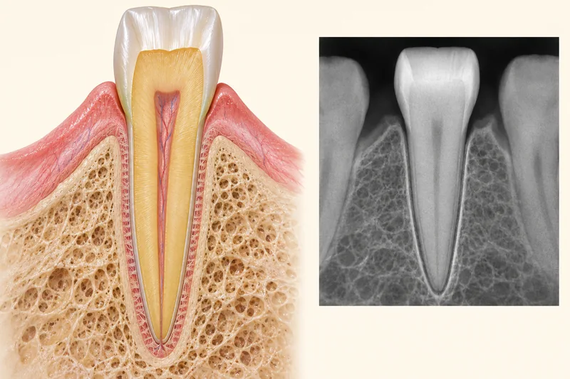
The root is not directly fused to the surrounding socket wall. The periodontal ligament lies between them. This anatomical space is not a hole inside the tooth.
The periodontal ligament space is more radiolucent than adjacent hard tissues. It may therefore appear dark beside the brighter socket boundary known as the lamina dura.
Width is not identical at every point. Projection and image quality also affect its appearance. An unusual finding is assessed alongside symptoms, examination and other findings.
The root is not directly fused to the surrounding socket wall. The periodontal ligament lies between them. This anatomical space is not a hole inside the tooth.
The periodontal ligament space is more radiolucent than adjacent hard tissues. It may therefore appear dark beside the brighter socket boundary known as the lamina dura.
Width is not identical at every point. Projection and image quality also affect its appearance. An unusual finding is assessed alongside symptoms, examination and other findings.
Yes, it belongs to the tooth-supporting tissues.
No. Radiographic contrast reflects X-ray attenuation.
No. Imaging and examination need to be combined.
Three steps. No audio.
Each scene can also be selected manually.

Silent educational film with German on-screen text and simplified teaching images. Not a patient case or a promised treatment outcome. The full English explanation is available as text.
The periodontal ligament space lies between root and bone. A periodontal pocket is examined from the gum margin and is not another name for this space around the root.
No. It contains the periodontal ligament between the root and socket. A dark line on a radiograph does not mean there is an empty cavity.
Unusual widening may merit closer assessment, but it does not establish a diagnosis by itself. Projection, the rest of the image and clinical findings need to be considered together.
No. A periodontal pocket extends from the gum margin alongside the tooth. The periodontal ligament space surrounds the root within the socket. They are different structures.
Not automatically. Symptoms, examination and existing images are considered first. An additional image is taken only when justified for a specific clinical question.
You can bring this question to your appointment:
Is the appearance at this tooth unusual, and how does it relate to the examination?
An examination can help clarify biting pain or questions about a radiographic finding.
Arrange an assessmentAppointment selection at DoctolibThe periodontal ligament space lies between root and bone. A periodontal pocket is examined from the gum margin and is not another name for this space around the root.
Editorial research: . General information, not an individual diagnosis or treatment recommendation. Teaching illustrations are simplified and do not depict a patient case.