Where does the tube go?
Into the natural opening in the mouth.

In conventional sialography a specialist introduces contrast dye through the natural opening of a major salivary duct. X-ray images then show the duct and its branches.
For training & practice ↓
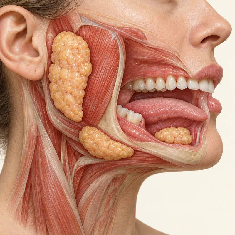
The parotid duct opens inside the cheek opposite the upper molars. The submandibular duct opens at the front of the floor of the mouth.
A small tube sits in the duct opening. Contrast dye passes through it and outlines the branches. This may cause a temporary feeling of pressure.
The images help look for abnormalities in the ducts, such as narrowing. The choice of examination depends on symptoms and previous findings.
The parotid duct opens inside the cheek opposite the upper molars. The submandibular duct opens at the front of the floor of the mouth.
A small tube sits in the duct opening. Contrast dye passes through it and outlines the branches. This may cause a temporary feeling of pressure.
The images help look for abnormalities in the ducts, such as narrowing. The choice of examination depends on symptoms and previous findings.
Into the natural opening in the mouth.
Contrast X-rays outline the ducts.
Tell the team about possible pregnancy or previous reactions to X-ray contrast.
Three steps. No audio.
Each scene can also be selected manually.

Silent film with English on-screen text and simplified teaching images.
A fine endoscope views the duct from inside. A sialogram is an X-ray image.
In a conventional sialogram it enters the salivary ducts through their opening.
The specialist chooses the test to suit the question. Ultrasound is often an initial investigation.
Contact the team about fever, acute pain or new swelling so they can advise about the examination.
Discussing imaging
What question should a sialogram answer in my case?
Seek medical or dental assessment for recurrent swelling to determine which investigation is appropriate.
Choose an appointmentAppointment selection at DoctolibThe appropriate test depends on whether the gland tissue, duct outline or inside of the duct needs assessing.
Editorial research: . General information, not an individual diagnosis or treatment recommendation. Teaching illustrations are simplified and do not depict a patient case.
If you turn this on, this website stores which guides you open, separately from advertising preferences. The markers expire after a fixed 30-day period from activation. The list is not sent to us by this feature. You can turn it off here and delete the markers.
Other people using the same browser profile can also see the markers. German and English versions share one marker. Expired entries are removed the next time you open a page with this feature; deleting browser data may remove them earlier. “Viewed” means opened, not read or completed.